 Français
 English

| ELK.No | ES3935 |
| Product name | CEACAM1/5 rabbit pAb |
| Reactivity | Human;Rat;Mouse; |
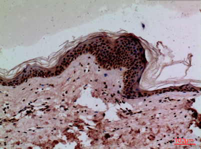
| Applications | WB;IHC;IF;ELISA |
| Other name | CEACAM1; BGP; BGP1; Carcinoembryonic antigen-related cell adhesion molecule 1; Biliary glycoprotein 1; BGP-1; CD66a; CEACAM5; CEA; Carcinoembryonic antigen-related cell adhesion molecule 5; Carcinoembryonic antigen; CEA; Meconium antigen 100; CD66e |
| Size | 50μL |
| Unit price ($) | 148 |
| Human gene ID | 634 |
| Human Swiss-Prot | P13688 |
| Source | Rabbit |
| Isotype | IgG |
| Target | CEACAM1/5 |
| Fields | |
| Gene name | CEACAM1/CEACAM5 |
| Protein name | Carcinoembryonic antigen-related cell adhesion molecule 1/Carcinoembryonic antigen-related cell adhesion molecule 5 |
| Human gene link | View Human Gene |
| Human Swiss link | View Human Swiss-Prot |
| Mouse gene ID | |
| Mouse gene link | |
| Mouse Swiss-Prot | P31809 |
| Mouse Swiss link | View Mouse Swiss-Prot |
| Rat gene ID | |
| Rat gene link | |
| Rat Swiss-Prot | |
| Rat Swiss link | |
| Immunogen | The antiserum was produced against synthesized peptide derived from the N-terminal region of human CEACAM1/CEACAM5. AA range:31-80 |
| Specificity | CEACAM1/5 Polyclonal Antibody detects endogenous levels of CEACAM1/5 protein. |
| Formulation | Liquid in PBS containing 50% glycerol, 0.5% BSA and 0.02% sodium azide. |
| Clonality | Polyclonal |
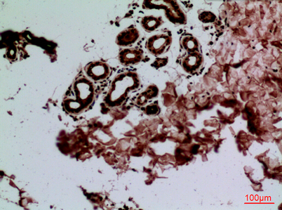
| Dilution | Western Blot: 1/500 - 1/2000. IHC-p: 1:100-300 ELISA: 1/20000. Not yet tested in other applications. |
| Purification | The antibody was affinity-purified from rabbit antiserum by affinity-chromatography using epitope-specific immunogen. |
| Concentration | 1 mg/ml |
| Storage stability | -20°C/1 year |
| Molecular Weight (Da) | |
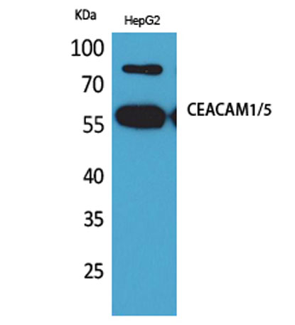
| Observed band (KD) | 58kD |
| Background | This gene encodes a member of the carcinoembryonic antigen (CEA) gene family, which belongs to the immunoglobulin superfamily. Two subgroups of the CEA family, the CEA cell adhesion molecules and the pregnancy-specific glycoproteins, are located within a 1.2 Mb cluster on the long arm of chromosome 19. Eleven pseudogenes of the CEA cell adhesion molecule subgroup are also found in the cluster. The encoded protein was originally described in bile ducts of liver as biliary glycoprotein. Subsequently, it was found to be a cell-cell adhesion molecule detected on leukocytes, epithelia, and endothelia. The encoded protein mediates cell adhesion via homophilic as well as heterophilic binding to other proteins of the subgroup. Multiple cellular activities have been attributed to the encoded protein, including roles in the differentiation and arrangement of tissue thre |
| Function | disease:Increased serum levels of BGP-1 are found in individuals suffering from hepatic disorders.,disease:Loss or reduced expression is a major event in colorectal carcinogenesis.,similarity:Belongs to the immunoglobulin superfamily. CEA family.,similarity:Contains 1 Ig-like V-type (immunoglobulin-like) domain.,similarity:Contains 3 Ig-like C2-type (immunoglobulin-like) domains., |
| Subcellular location | [Isoform 1]: Cell membrane ; Single-pass type I membrane protein . Lateral cell membrane . Apical cell membrane . Basal cell membrane . Cell junction . Cell junction, adherens junction . Canalicular domain of hepatocyte plasma membranes. Found as a mixture of monomer, dimer and oligomer in the plasma membrane. Occurs predominantly as cis-dimers and/or small cis-oligomers in the cell junction regions. Found as dimer in the solution. Predominantly localized to the lateral cell membranes. .; [Isoform 2]: Secreted .; [Isoform 3]: Secreted .; [Isoform 4]: Secreted .; [Isoform 5]: Cell membrane; Single-pass type I membrane protein.; [Isoform 6]: Cell membrane; Single-pass type I membrane protein.; [Isoform 7]: Cell membrane; Single-pass type I membrane protein.; [Isoform 8]: Cell membrane ; Sing |
| Expression | Expressed in columnar epithelial cells of the colon (at protein level) (PubMed:10436421). The predominant forms expressed by T cells are those containing a long cytoplasmic domain (PubMed:18424730). Expressed in granulocytes and lymphocytes. Leukocytes only express isoforms 6 and isoform 1 (PubMed:11994468). |

Use collapsible tabs for more detailed information that will help customers make a purchasing decision.
Ex: Shipping and return policies, size guides, and other common questions.