 Français
 English

| ELK.No | ES3968 |
| Product name | CD1C rabbit pAb |
| Reactivity | Human;Rat;Mouse; |
| Applications | WB;ELISA |
| Other name | CD1C; T-cell surface glycoprotein CD1c; CD1c |
| Size | 50μL |
| Unit price ($) | 148 |
| Human gene ID | 911 |
| Human Swiss-Prot | P29017 |
| Source | Rabbit |
| Isotype | IgG |
| Target | CD1C |
| Fields | >>Tight junction;>>Hematopoietic cell lineage;>>Amoebiasis |
| Gene name | CD1C |
| Protein name | T-cell surface glycoprotein CD1c |
| Human gene link | View Human Gene |
| Human Swiss link | View Human Swiss-Prot |
| Mouse gene ID | |
| Mouse gene link | |
| Mouse Swiss-Prot | |
| Mouse Swiss link | |
| Rat gene ID | |
| Rat gene link | |
| Rat Swiss-Prot | |
| Rat Swiss link | |
| Immunogen | The antiserum was produced against synthesized peptide derived from the Internal region of human CD1C. AA range:211-260 |
| Specificity | CD1C Polyclonal Antibody detects endogenous levels of CD1C protein. |
| Formulation | Liquid in PBS containing 50% glycerol, 0.5% BSA and 0.02% sodium azide. |
| Clonality | Polyclonal |
| Dilution | Western Blot: 1/500 - 1/2000. ELISA: 1/20000. Not yet tested in other applications. |
| Purification | The antibody was affinity-purified from rabbit antiserum by affinity-chromatography using epitope-specific immunogen. |
| Concentration | 1 mg/ml |
| Storage stability | -20°C/1 year |
| Molecular Weight (Da) | |
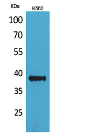
| Observed band (KD) | 37kD |
| Background | This gene encodes a member of the CD1 family of transmembrane glycoproteins, which are structurally related to the major histocompatibility complex (MHC) proteins and form heterodimers with beta-2-microglobulin. The CD1 proteins mediate the presentation of primarily lipid and glycolipid antigens of self or microbial origin to T cells. The human genome contains five CD1 family genes organized in a cluster on chromosome 1. The CD1 family members are thought to differ in their cellular localization and specificity for particular lipid ligands. The protein encoded by this gene is broadly distributed throughout the endocytic system via a tyrosine-based motif in the cytoplasmic tail. Alternatively spliced transcript variants of this gene have been observed, but their full-length nature is not known. [provided by RefSeq, Jul 2008], |
| Function | function:Antigen-presenting protein that binds self and non-self lipid and glycolipid antigens and presents them to T-cell receptors on natural killer T-cells.,miscellaneous:During protein synthesis and maturation, CD1 family members bind endogenous lipids that are replaced by lipid or glycolipid antigens when the proteins are internalized and pass through endosomes or lysosomes, before trafficking back to the cell surface.,similarity:Contains 1 Ig-like (immunoglobulin-like) domain.,subcellular location:Subject to intracellular trafficking between the cell membrane and endosomes.,subunit:Heterodimer with B2M (beta-2-microglobulin).,tissue specificity:Expressed on cortical thymocytes, on certain T-cell leukemias, and in various other tissues., |
| Subcellular location | Cell membrane ; Single-pass type I membrane protein . Endosome membrane ; Single-pass type I membrane protein. Lysosome . Subject to intracellular trafficking between the cell membrane and endosomes. . |
| Expression | Expressed on cortical thymocytes, on certain T-cell leukemias, and in various other tissues. |

Use collapsible tabs for more detailed information that will help customers make a purchasing decision.
Ex: Shipping and return policies, size guides, and other common questions.