 Français
 English
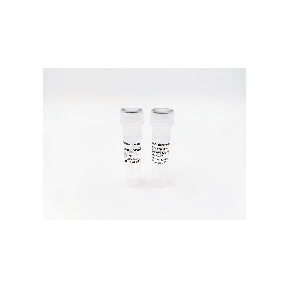

| ELK.No | ES4011 |
| Product name | CD328 rabbit pAb |
| Reactivity | Human;Rat;Mouse; |
| Applications | WB;IHC;IF;ELISA |
| Other name | SIGLEC7; AIRM1; Sialic acid-binding Ig-like lectin 7; Siglec-7; Adhesion inhibitory receptor molecule 1; AIRM-1; CDw328; D-siglec; QA79 membrane protein; p75; CD328 |
| Size | 50μL |
| Unit price ($) | 148 |
| Human gene ID | 27036 |
| Human Swiss-Prot | Q9Y286 |
| Source | Rabbit |
| Isotype | IgG |
| Target | CD328 |
| Fields | |
| Gene name | SIGLEC7 |
| Protein name | Sialic acid-binding Ig-like lectin 7 |
| Human gene link | View Human Gene |
| Human Swiss link | View Human Swiss-Prot |
| Mouse gene ID | |
| Mouse gene link | |
| Mouse Swiss-Prot | |
| Mouse Swiss link | |
| Rat gene ID | |
| Rat gene link | |
| Rat Swiss-Prot | |
| Rat Swiss link | |
| Immunogen | The antiserum was produced against synthesized peptide derived from the Internal region of human SIGLEC7. AA range:51-100 |
| Specificity | CD328 Polyclonal Antibody detects endogenous levels of CD328 protein. |
| Formulation | Liquid in PBS containing 50% glycerol, 0.5% BSA and 0.02% sodium azide. |
| Clonality | Polyclonal |
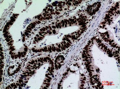
| Dilution | Western Blot: 1/500 - 1/2000. IHC-p: 1:100-300 ELISA: 1/20000. Not yet tested in other applications. |
| Purification | The antibody was affinity-purified from rabbit antiserum by affinity-chromatography using epitope-specific immunogen. |
| Concentration | 1 mg/ml |
| Storage stability | -20°C/1 year |
| Molecular Weight (Da) | |
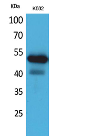
| Observed band (KD) | 51kD |
| Background | domain:Contains 1 copy of a cytoplasmic motif that is referred to as the immunoreceptor tyrosine-based inhibitor motif (ITIM). This motif is involved in modulation of cellular responses. The phosphorylated ITIM motif can bind the SH2 domain of several SH2-containing phosphatases.,function:Putative adhesion molecule that mediates sialic-acid dependent binding to cells. Preferentially binds to alpha-2,3- and alpha-2,6-linked sialic acid. Also binds disialogangliosides (disialogalactosyl globoside, disialyl lactotetraosylceramide and disialyl GalNAc lactotetraoslylceramide). The sialic acid recognition site may be masked by cis interactions with sialic acids on the same cell surface. In the immune response, may act as an inhibitory receptor upon ligand induced tyrosine phosphorylation by recruiting cytoplasmic phosphatase(s) via their SH2 domain(s) that block signal transduction through dephosphorylation of signaling molecules. Mediates inhibition of natural killer cells cytotoxicity. May play a role in hemopoiesis. Inhibits differentiation of CD34+ cell precursors towards myelomonocytic cell lineage and proliferation of leukemic myeloid cells (in vitro).,online information:Siglec-7,PTM:Tyrosine phosphorylated.,similarity:Belongs to the immunoglobulin superfamily. SIGLEC (sialic acid binding Ig-like lectin) family.,similarity:Contains 1 Ig-like V-type (immunoglobulin-like) domain.,similarity:Contains 2 Ig-like C2-type (immunoglobulin-like) domains.,subunit:Interacts with PTPN6/SHP-1 upon phosphorylation.,tissue specificity:Predominantly expressed by resting and activated natural killer cells and at lower levels by granulocytes and monocytes. High expression found in placenta, liver, lung, spleen, and peripheral blood leukocytes., |
| Function | domain:Contains 1 copy of a cytoplasmic motif that is referred to as the immunoreceptor tyrosine-based inhibitor motif (ITIM). This motif is involved in modulation of cellular responses. The phosphorylated ITIM motif can bind the SH2 domain of several SH2-containing phosphatases.,function:Putative adhesion molecule that mediates sialic-acid dependent binding to cells. Preferentially binds to alpha-2,3- and alpha-2,6-linked sialic acid. Also binds disialogangliosides (disialogalactosyl globoside, disialyl lactotetraosylceramide and disialyl GalNAc lactotetraoslylceramide). The sialic acid recognition site may be masked by cis interactions with sialic acids on the same cell surface. In the immune response, may act as an inhibitory receptor upon ligand induced tyrosine phosphorylation by recruiting cytoplasmic phosphatase(s) via their SH2 domain(s) that block signal transduction through dep |
| Subcellular location | Membrane; Single-pass type I membrane protein. |
| Expression | Predominantly expressed by resting and activated natural killer cells and at lower levels by granulocytes and monocytes. High expression found in placenta, liver, lung, spleen, and peripheral blood leukocytes. |


Use collapsible tabs for more detailed information that will help customers make a purchasing decision.
Ex: Shipping and return policies, size guides, and other common questions.