 Français
 English

| ELK.No | ES4046 |
| Product name | Amphiphysin II rabbit pAb |
| Reactivity | Mouse;Rat |
| Applications | WB;IHC;IF;ELISA |
| Other name | BIN1; AMPHL; Myc box-dependent-interacting protein 1; Amphiphysin II; Amphiphysin-like protein; Box-dependent myc-interacting protein 1; Bridging integrator 1 |
| Size | 50μL |
| Unit price ($) | 148 |
| Human gene ID | 274 |
| Human Swiss-Prot | O00499 |
| Source | Rabbit |
| Isotype | IgG |
| Target | Amphiphysin II |
| Fields | >>Endocytosis;>>Fc gamma R-mediated phagocytosis |
| Gene name | BIN1 |
| Protein name | Myc box-dependent-interacting protein 1 |
| Human gene link | View Human Gene |
| Human Swiss link | View Human Swiss-Prot |
| Mouse gene ID | 30948 |
| Mouse gene link | View Mouse Gene |
| Mouse Swiss-Prot | O08539 |
| Mouse Swiss link | View Mouse Swiss-Prot |
| Rat gene ID | 117028 |
| Rat gene link | View Rat Gene |
| Rat Swiss-Prot | O08839 |
| Rat Swiss link | View Rat Swiss-Prot |
| Immunogen | Synthesized peptide derived from the C-terminal region of human Amphiphysin II. |
| Specificity | Amphiphysin II Polyclonal Antibody detects endogenous levels of Amphiphysin II protein. |
| Formulation | Liquid in PBS containing 50% glycerol, 0.5% BSA and 0.02% sodium azide. |
| Clonality | Polyclonal |
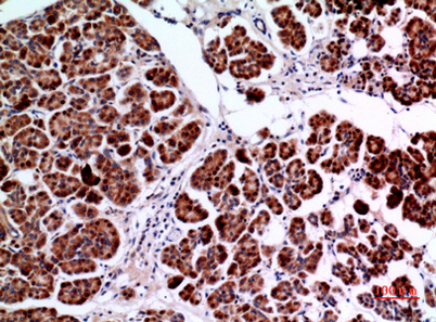
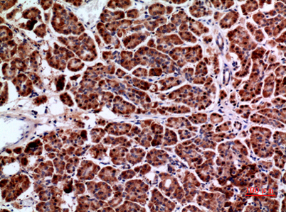
| Dilution | Western Blot: 1/500 - 1/2000. IHC-p: 1/100-1/300. ELISA: 1/20000. Not yet tested in other applications. |
| Purification | The antibody was affinity-purified from rabbit antiserum by affinity-chromatography using epitope-specific immunogen. |
| Concentration | 1 mg/ml |
| Storage stability | -20°C/1 year |
| Molecular Weight (Da) | |
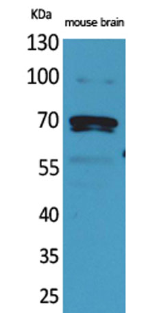
| Observed band (KD) | 64kD |
| Background | This gene encodes several isoforms of a nucleocytoplasmic adaptor protein, one of which was initially identified as a MYC-interacting protein with features of a tumor suppressor. Isoforms that are expressed in the central nervous system may be involved in synaptic vesicle endocytosis and may interact with dynamin, synaptojanin, endophilin, and clathrin. Isoforms that are expressed in muscle and ubiquitously expressed isoforms localize to the cytoplasm and nucleus and activate a caspase-independent apoptotic process. Studies in mouse suggest that this gene plays an important role in cardiac muscle development. Alternate splicing of the gene results in several transcript variants encoding different isoforms. Aberrant splice variants expressed in tumor cell lines have also been described. [provided by RefSeq, Mar 2016], |
| Function | alternative products:Additional isoforms seem to exist,disease:Defects in BIN1 are the cause of centronuclear myopathy autosomal recessive (ARCNM) [MIM:255200]; also known as autosomal recessive myotubular myopathy. Centronuclear myopathies are congenital muscle disorders characterized by progressive muscular weakness and wasting involving mainly limb girdle, trunk, and neck muscles. It may also affect distal muscles. Weakness may be present during childhood or adolescence or may not become evident until the third decade of life. Ptosis is a frequent clinical feature. The most prominent histopathologic features include high frequency of centrally located nuclei in muscle fibers not secondary to regeneration, radial arrangement of sarcoplasmic strands around the central nuclei, and predominance and hypotrophy of type 1 fibers.,function:May be involved in regulation of synaptic vesicle end |
| Subcellular location | [Isoform BIN1]: Nucleus . Cytoplasm . Endosome . Cell membrane, sarcolemma, T-tubule .; [Isoform IIA]: Cytoplasm . |
| Expression | Ubiquitous. Highest expression in the brain and muscle (PubMed:9182667). Expressed in oligodendrocytes (PubMed:27488240). Isoform IIA is expressed only in the brain, where it is detected in the gray matter, but not in the white matter (PubMed:27488240). Isoform BIN1 is widely expressed with highest expression in skeletal muscle. |

Use collapsible tabs for more detailed information that will help customers make a purchasing decision.
Ex: Shipping and return policies, size guides, and other common questions.