 Français
 English

| ELK.No | ES4055 |
| Product name | EPAS-1 rabbit pAb |
| Reactivity | Human;Mouse;Rat |
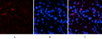
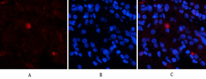
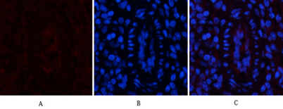
| Applications | IF;WB;IHC;ELISA |
| Other name | EPAS1; BHLHE73; HIF2A; MOP2; PASD2; Endothelial PAS domain-containing protein 1; EPAS-1; Basic-helix-loop-helix-PAS protein MOP2; Class E basic helix-loop-helix protein 73; bHLHe73;HIF-1-alpha-like factor; HLF; Hypoxia-inducible factor 2-alpha; HIF-2-alpha; HIF2-alpha; Member of PAS protein 2; PAS domain-containing protein 2 |
| Size | 50μL |
| Unit price ($) | 148 |
| Human gene ID | 2034 |
| Human Swiss-Prot | Q99814 |
| Source | Rabbit |
| Isotype | IgG |
| Target | EPAS-1 |
| Fields | >>Pathways in cancer;>>Renal cell carcinoma |
| Gene name | EPAS1 BHLHE73 HIF2A MOP2 PASD2 |
| Protein name | Endothelial PAS domain-containing protein 1 |
| Human gene link | View Human Gene |
| Human Swiss link | View Human Swiss-Prot |
| Mouse gene ID | 13819 |
| Mouse gene link | View Mouse Gene |
| Mouse Swiss-Prot | P97481 |
| Mouse Swiss link | View Mouse Swiss-Prot |
| Rat gene ID | 29452 |
| Rat gene link | View Rat Gene |
| Rat Swiss-Prot | Q9JHS1 |
| Rat Swiss link | View Rat Swiss-Prot |
| Immunogen | Synthesized peptide derived from human EPAS-1 around the non-acetylation site of K385. |
| Specificity | EPAS-1 Polyclonal Antibody detects endogenous levels of EPAS-1 protein. |
| Formulation | Liquid in PBS containing 50% glycerol, 0.5% BSA and 0.02% sodium azide. |
| Clonality | Polyclonal |
| Dilution | IF: 1:50-200 Western Blot: 1/500 - 1/2000. IHC-p: 1/100-1/300. ELISA: 1/20000. Not yet tested in other applications. |
| Purification | The antibody was affinity-purified from rabbit antiserum by affinity-chromatography using epitope-specific immunogen. |
| Concentration | 1 mg/ml |
| Storage stability | -20°C/1 year |
| Molecular Weight (Da) | |
| Observed band (KD) | 96kD |
| Background | endothelial PAS domain protein 1(EPAS1) Homo sapiens This gene encodes a transcription factor involved in the induction of genes regulated by oxygen, which is induced as oxygen levels fall. The encoded protein contains a basic-helix-loop-helix domain protein dimerization domain as well as a domain found in proteins in signal transduction pathways which respond to oxygen levels. Mutations in this gene are associated with erythrocytosis familial type 4. [provided by RefSeq, Nov 2009], |
| Function | disease:Defects in EPAS1 are the cause of erythrocytosis familial type 4 (ECYT4) [MIM:611783]. ECYT4 is an autosomal dominant disorder characterized by increased serum red blood cell mass, elevated hemoglobin concentration and hematocrit, and normal platelet and leukocyte counts.,function:Transcription factor involved in the induction of oxygen regulated genes. Binds to core DNA sequence 5'-[AG]CGTG-3' within the hypoxia response element (HRE) of target gene promoters. Regulates the vascular endothelial growth factor (VEGF) expression and seems to be implicated in the development of blood vessels and the tubular system of lung. May also play a role in the formation of the endothelium that gives rise to the blood brain barrier. Potent activator of the Tie-2 tyrosine kinase expression. Activation seems to require recruitment of transcriptional coactivators such as CREBPB and probably EP300 |
| Subcellular location | Nucleus . Nucleus speckle . Colocalizes with HIF3A in the nucleus and speckles. . |
| Expression | Expressed in most tissues, with highest levels in placenta, lung and heart. Selectively expressed in endothelial cells. |

Use collapsible tabs for more detailed information that will help customers make a purchasing decision.
Ex: Shipping and return policies, size guides, and other common questions.