 Français
 English
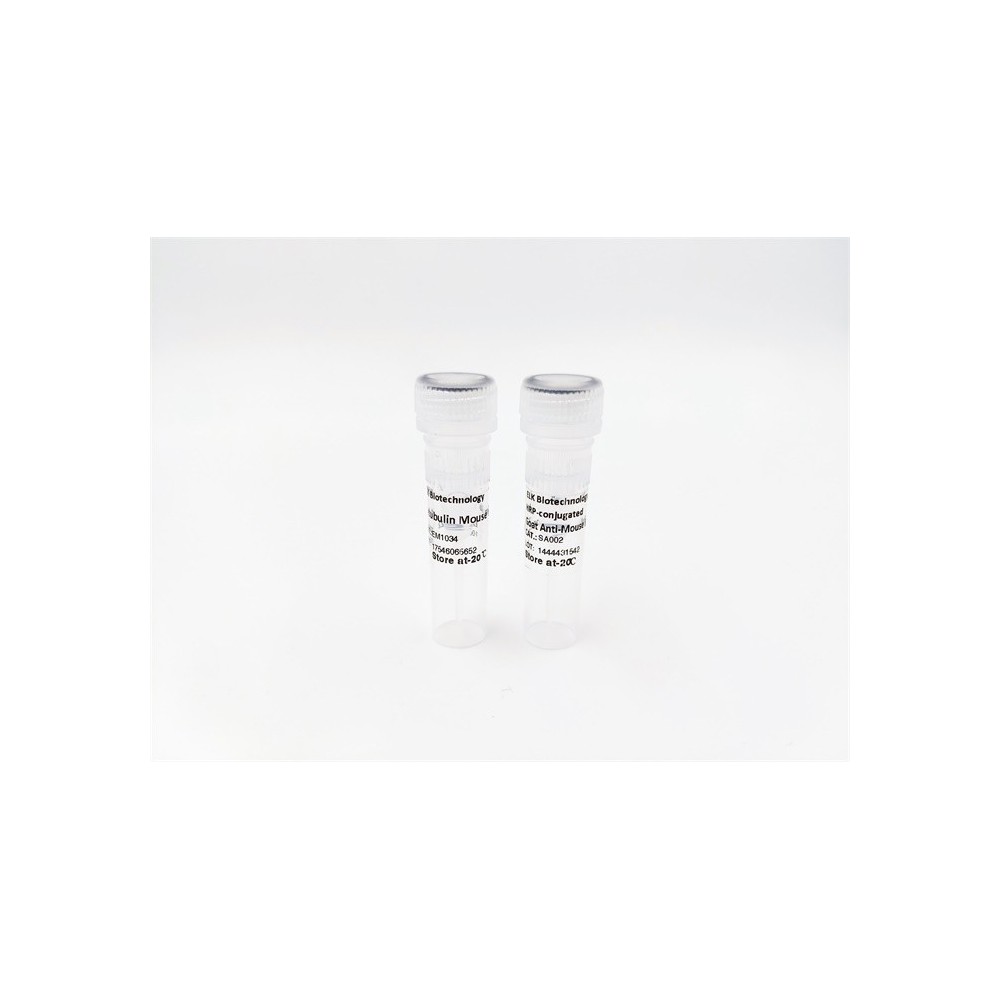

| ELK.No | ES4112 |
| Product name | Cryopyrin rabbit pAb |
| Reactivity | Human;Mouse;Rat |
| Applications | WB;IHC;IF;ELISA |
| Other name | NLRP3; C1orf7; CIAS1; NALP3; PYPAF1; NACHT, LRR and PYD domains-containing protein 3; Angiotensin/vasopressin receptor AII/AVP-like; Caterpiller protein 1.1CLR1.1; Cold autoinflammatory syndrome 1 protein; Cryopyrin; PYRIN-containing APAF1-like protein 1 |
| Size | 100μL |
| Unit price ($) | 248 |
| Human gene ID | 114548 |
| Human Swiss-Prot | Q96P20 |
| Source | Rabbit |
| Isotype | IgG |
| Target | Cryopyrin |
| Fields | >>Necroptosis;>>NOD-like receptor signaling pathway;>>C-type lectin receptor signaling pathway;>>Pathogenic Escherichia coli infection;>>Shigellosis;>>Salmonella infection;>>Pertussis;>>Yersinia infection;>>Influenza A;>>Coronavirus disease - COVID-19;>>Lipid and atherosclerosis |
| Gene name | NLRP3 |
| Protein name | NACHT LRR and PYD domains-containing protein 3 |
| Human gene link | View Human Gene |
| Human Swiss link | View Human Swiss-Prot |
| Mouse gene ID | |
| Mouse gene link | |
| Mouse Swiss-Prot | Q8R4B8 |
| Mouse Swiss link | View Mouse Swiss-Prot |
| Rat gene ID | |
| Rat gene link | |
| Rat Swiss-Prot | |
| Rat Swiss link | |
| Immunogen | The antiserum was produced against synthesized peptide derived from the Internal region of human NLRP3. AA range:511-560 |
| Specificity | Cryopyrin Polyclonal Antibody detects endogenous levels of Cryopyrin protein. |
| Formulation | Liquid in PBS containing 50% glycerol, 0.5% BSA and 0.02% sodium azide. |
| Clonality | Polyclonal |
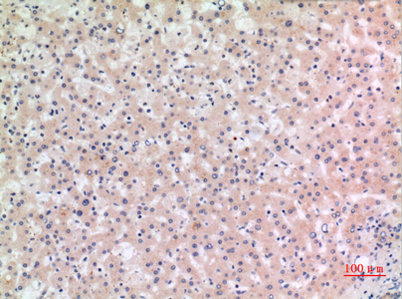
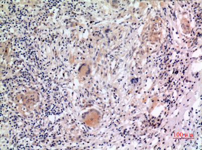
| Dilution | Western Blot: 1/500 - 1/2000. IHC-p: 1/100-1/300. ELISA: 1/20000. Not yet tested in other applications. |
| Purification | The antibody was affinity-purified from rabbit antiserum by affinity-chromatography using epitope-specific immunogen. |
| Concentration | 1 mg/ml |
| Storage stability | -20°C/1 year |
| Molecular Weight (Da) | |
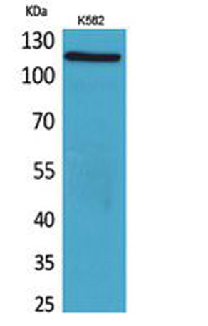
| Observed band (KD) | 115kD |
| Background | This gene encodes a pyrin-like protein containing a pyrin domain, a nucleotide-binding site (NBS) domain, and a leucine-rich repeat (LRR) motif. This protein interacts with the apoptosis-associated speck-like protein PYCARD/ASC, which contains a caspase recruitment domain, and is a member of the NALP3 inflammasome complex. This complex functions as an upstream activator of NF-kappaB signaling, and it plays a role in the regulation of inflammation, the immune response, and apoptosis. Mutations in this gene are associated with familial cold autoinflammatory syndrome (FCAS), Muckle-Wells syndrome (MWS), chronic infantile neurological cutaneous and articular (CINCA) syndrome, and neonatal-onset multisystem inflammatory disease (NOMID). Multiple alternatively spliced transcript variants encoding distinct isoforms have been identified for this gene. Alternative 5' UTR structures are s |
| Function | disease:Defects in NLRP3 are a cause of Muckle-Wells syndrome (MWS) [MIM:191900]; also known as urticaria-deafness-amyloidosis syndrome. MWS is a hereditary periodic fever syndrome characterized by fever, chronic recurrent urticaria, arthralgias, progressive sensorineural deafness, and reactive renal amyloidosis. The disease may be severe if generalized amyloidosis occurs.,disease:Defects in NLRP3 are the cause of chronic infantile neurologic cutaneous and articular syndrome (CINCA) [MIM:607115]; also known as 'neonatal onset multisystem inflammatory disease,' or NOMID, a rare congenital inflammatory disorder characterized by a triad of neonatal onset of cutaneous symptoms, chronic meningitis, and joint manifestations with recurrent fever and inflammation.,disease:Defects in NLRP3 are the cause of familial cold autoinflammatory syndrome type 1 (FCAS1) [MIM:120100]; commonly known as fami |
| Subcellular location | Cytoplasm, cytosol . Inflammasome . Endoplasmic reticulum . Secreted . Nucleus . In macrophages, under resting conditions, mainly located in the cytosol, on the endoplasmic reticulum. After stimulation with inducers of the NLRP3 inflammasome, mitochondria redistribute in the vicinity of the endoplasmic reticulum in the perinuclear region, which results in colocalization of NLRP3 on the endoplasmic reticulum and PYCARD on mitochondria, allowing the activation of inflammasome assembly. After the induction of pyroptosis, inflammasome specks are released into the extracellular space where they can further promote IL1B processing and where they can be engulfed by macrophages. Phagocytosis induces lysosomal damage and inflammasome activation in the recipient cells (PubMed:24952504). In the Th2 s |
| Expression | Predominantly expressed in macrophages (PubMed:33231615, PubMed:34133077). Also expressed in dendritic cells, B- and T-cells (at protein level) (PubMed:11786556) (PubMed:17164409). Expressed in LPS-treated granulocytes, but not in resting cells (at protein level) (PubMed:17164409). Expression in monocytes is very weak (at protein level) (PubMed:17164409). Expressed in stratified non-keratinizing squamous epithelium, including oral, esophageal and ectocervical mucosa and in the Hassall's corpuscles in the thymus. Also, detected in the stratified epithelium covering the bladder and ureter (transitional mucosa) (at protein level) (PubMed:17164409). Expressed in lung epithelial cells (at protein level) (PubMed:23229815). Expressed in chondrocytes (PubMed:12032915). Expressed at low levels in r |

Use collapsible tabs for more detailed information that will help customers make a purchasing decision.
Ex: Shipping and return policies, size guides, and other common questions.