 Français
 English
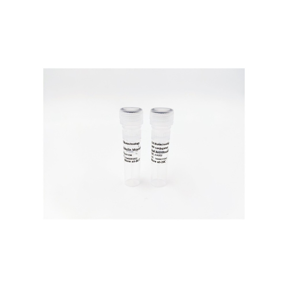

| ELK.No | ES4196 |
| Product name | LIR-7 rabbit pAb |
| Reactivity | Human;Rat;Mouse; |
| Applications | WB;IHC;IF;ELISA |
| Other name | LILRA2; ILT1; LIR7; Leukocyte immunoglobulin-like receptor subfamily A member 2; CD85 antigen-like family member H; Immunoglobulin-like transcript 1; ILT-1; Leukocyte immunoglobulin-like receptor 7; LIR-7; CD85h |
| Size | 50μL |
| Unit price ($) | 148 |
| Human gene ID | 11027 |
| Human Swiss-Prot | Q8N149 |
| Source | Rabbit |
| Isotype | IgG |
| Target | LIR-7 |
| Fields | >>Osteoclast differentiation;>>B cell receptor signaling pathway |
| Gene name | LILRA2 |
| Protein name | Leukocyte immunoglobulin-like receptor subfamily A member 2 |
| Human gene link | View Human Gene |
| Human Swiss link | View Human Swiss-Prot |
| Mouse gene ID | |
| Mouse gene link | |
| Mouse Swiss-Prot | |
| Mouse Swiss link | |
| Rat gene ID | |
| Rat gene link | |
| Rat Swiss-Prot | |
| Rat Swiss link | |
| Immunogen | The antiserum was produced against synthesized peptide derived from the Internal region of human LILRA2. AA range:121-170 |
| Specificity | LIR-7 Polyclonal Antibody detects endogenous levels of LIR-7 protein. |
| Formulation | Liquid in PBS containing 50% glycerol, 0.5% BSA and 0.02% sodium azide. |
| Clonality | Polyclonal |
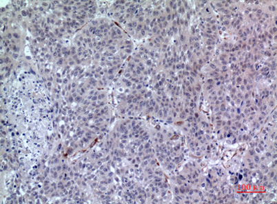
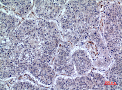
| Dilution | Western Blot: 1/500 - 1/2000. IHC-p: 1/100-1/300. ELISA: 1/20000. Not yet tested in other applications. |
| Purification | The antibody was affinity-purified from rabbit antiserum by affinity-chromatography using epitope-specific immunogen. |
| Concentration | 1 mg/ml |
| Storage stability | -20°C/1 year |
| Molecular Weight (Da) | |
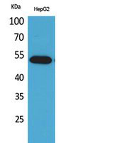
| Observed band (KD) | 53kD |
| Background | This gene encodes a member of a family of immunoreceptors that are expressed predominantly on monocytes and B cells, and at lower levels on dendritic cells and natural killer cells. The encoded protein is an activating receptor that inhibits dendritic cell differentiation and antigen presentation and suppresses innate immune response. Alternatively spliced transcript variants encoding different isoforms have been found. This gene is located in a cluster of related genes on chromosome 19 and there is a pseudogene for this gene on chromosome 3. [provided by RefSeq, Mar 2014], |
| Function | function:May act as receptor for class I MHC antigens.,similarity:Contains 4 Ig-like C2-type (immunoglobulin-like) domains.,tissue specificity:Expression levels are very low or not detectable on monocytes, T-cells, B-cells, dendritic cells and natural killer (NK) cells., |
| Subcellular location | Cell membrane ; Single-pass type I membrane protein .; [Isoform 4]: Secreted . |
| Expression | Detected on the surface of all peripheral blood monocytes, neutrophils, basophils and eosinophils (at protein level) (PubMed:12529506, PubMed:22479404). Expression levels are very low or not detectable on monocytes, T-cells, B-cells, dendritic cells and natural killer (NK) cells (PubMed:9548455). |

Use collapsible tabs for more detailed information that will help customers make a purchasing decision.
Ex: Shipping and return policies, size guides, and other common questions.