 Français
 English

| ELK.No | ES4242 |
| Product name | IL-1F8 rabbit pAb |
| Reactivity | Human;Rat;Mouse; |
| Applications | WB;IHC;IF;ELISA |
| Other name | IL36B; IL1F8; IL1H2; Interleukin-36 beta; FIL1 eta; Interleukin-1 eta; IL-1 eta; Interleukin-1 family member 8; IL-1F8; Interleukin-1 homolog 2; IL-1H2 |
| Size | 100μL |
| Unit price ($) | 248 |
| Human gene ID | 27177 |
| Human Swiss-Prot | Q9NZH7 |
| Source | Rabbit |
| Isotype | IgG |
| Target | IL-1F8 |
| Fields | >>Cytokine-cytokine receptor interaction |
| Gene name | IL36B |
| Protein name | Interleukin-36 beta |
| Human gene link | View Human Gene |
| Human Swiss link | View Human Swiss-Prot |
| Mouse gene ID | 69677 |
| Mouse gene link | View Mouse Gene |
| Mouse Swiss-Prot | Q9D6Z6 |
| Mouse Swiss link | View Mouse Swiss-Prot |
| Rat gene ID | |
| Rat gene link | |
| Rat Swiss-Prot | |
| Rat Swiss link | |
| Immunogen | The antiserum was produced against synthesized peptide derived from the C-terminal region of human IL36B. AA range:111-160 |
| Specificity | IL-1F8 Polyclonal Antibody detects endogenous levels of IL-1F8 protein. |
| Formulation | Liquid in PBS containing 50% glycerol, 0.5% BSA and 0.02% sodium azide. |
| Clonality | Polyclonal |
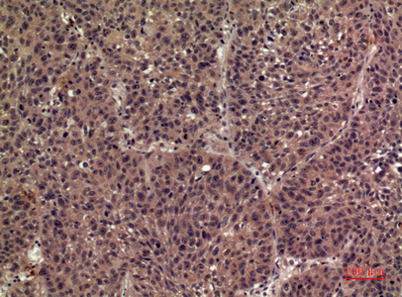
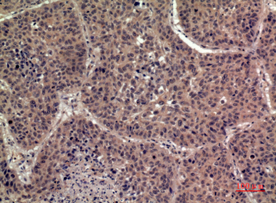
| Dilution | Western Blot: 1/500 - 1/2000. IHC-p: 1/100-1/300. ELISA: 1/20000. Not yet tested in other applications. |
| Purification | The antibody was affinity-purified from rabbit antiserum by affinity-chromatography using epitope-specific immunogen. |
| Concentration | 1 mg/ml |
| Storage stability | -20°C/1 year |
| Molecular Weight (Da) | |
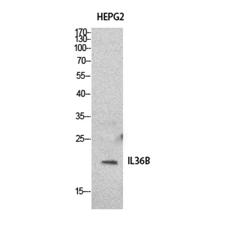
| Observed band (KD) | 19kD |
| Background | The protein encoded by this gene is a member of the interleukin 1 cytokine family. Protein structure modeling indicated that this cytokine may contain a 12-stranded beta-trefoil structure that is conserved between IL1A (IL-A alpha) and IL1B (IL-1 beta). This gene and eight other interleukin 1 family genes form a cytokine gene cluster on chromosome 2. Two alternatively spliced transcript variants encoding distinct isoforms have been reported. [provided by RefSeq, Jul 2008], |
| Function | online information:Interleukin-1 entry,similarity:Belongs to the IL-1 family.,tissue specificity:Expression at low levels in tonsil, bone marrow, heart, placenta, lung, testis and colon but not in other tissues or in any hematopoietic cell lines., |
| Subcellular location | Cytoplasm . Secreted . The secretion is dependent on protein unfolding and facilitated by the cargo receptor TMED10; it results in protein translocation from the cytoplasm into the ERGIC (endoplasmic reticulum-Golgi intermediate compartment) followed by vesicle entry and secretion. . |
| Expression | Expression at low levels in tonsil, bone marrow, heart, placenta, lung, testis and colon but not in any hematopoietic cell lines. Not detected in adipose tissue. Expressed at higher levels in psoriatic plaques than in symptomless psoriatic skin or healthy control skin. Increased levels are not detected in inflamed joint tissue. |

Use collapsible tabs for more detailed information that will help customers make a purchasing decision.
Ex: Shipping and return policies, size guides, and other common questions.