 Français
 English

| ELK.No | ES4255 |
| Product name | CD63 rabbit pAb |
| Reactivity | Human;Rat;Mouse; |
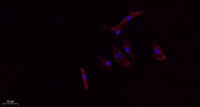
| Applications | IF;WB;IHC;ELISA |
| Other name | CD63; MLA1; TSPAN30; CD63 antigen; Granulophysin; Lysosomal-associated membrane protein 3; LAMP-3; Melanoma-associated antigen ME491; OMA81H; Ocular melanoma-associated antigen; Tetraspanin-30; Tspan-30; CD63 |
| Size | 100μL |
| Unit price ($) | 248 |
| Human gene ID | 967 |
| Human Swiss-Prot | P08962 |
| Source | Rabbit |
| Isotype | IgG |
| Target | CD63 |
| Fields | >>Lysosome;>>Proteoglycans in cancer |
| Gene name | CD63 |
| Protein name | CD63 antigen |
| Human gene link | View Human Gene |
| Human Swiss link | View Human Swiss-Prot |
| Mouse gene ID | |
| Mouse gene link | |
| Mouse Swiss-Prot | P41731 |
| Mouse Swiss link | View Mouse Swiss-Prot |
| Rat gene ID | |
| Rat gene link | |
| Rat Swiss-Prot | |
| Rat Swiss link | |
| Immunogen | The antiserum was produced against synthesized peptide derived from the Internal region of human CD63. AA range:121-170 |
| Specificity | CD63 Polyclonal Antibody detects endogenous levels of CD63 protein. |
| Formulation | Liquid in PBS containing 50% glycerol, 0.5% BSA and 0.02% sodium azide. |
| Clonality | Polyclonal |
| Dilution | IF: 1:50-200 Western Blot: 1/500 - 1/2000. IHC-p: 1:100-1:300. ELISA: 1/20000. Not yet tested in other applications. |
| Purification | The antibody was affinity-purified from rabbit antiserum by affinity-chromatography using epitope-specific immunogen. |
| Concentration | 1 mg/ml |
| Storage stability | -20°C/1 year |
| Molecular Weight (Da) | |
| Observed band (KD) | 26,35-65(kD |
| Background | The protein encoded by this gene is a member of the transmembrane 4 superfamily, also known as the tetraspanin family. Most of these members are cell-surface proteins that are characterized by the presence of four hydrophobic domains. The proteins mediate signal transduction events that play a role in the regulation of cell development, activation, growth and motility. The encoded protein is a cell surface glycoprotein that is known to complex with integrins. It may function as a blood platelet activation marker. Deficiency of this protein is associated with Hermansky-Pudlak syndrome. Also this gene has been associated with tumor progression. Alternative splicing results in multiple transcript variants encoding different protein isoforms. [provided by RefSeq, Apr 2012], |
| Function | function:This antigen is associated with early stages of melanoma tumor progression. May play a role in growth regulation.,miscellaneous:Lack of expression of CD63 in platelets has been observed in a patient with Hermansky-Pudlak syndrome (HPS). Hermansky-Pudlak syndrome (HPS) is a genetically heterogeneous, rare, autosomal recessive disorder characterized by oculocutaneous albinism, bleeding due to platelet storage pool deficiency, and lysosomal storage defects. This syndrome results from defects of diverse cytoplasmic organelles including melanosomes, platelet dense granules and lysosomes. Ceroid storage in the lungs is associated with pulmonary fibrosis, a common cause of premature death in individuals with HPS.,similarity:Belongs to the tetraspanin (TM4SF) family.,subcellular location:Also found in Weibel-Palade bodies of endothelial cells. Located in platelet dense granules.,tissue |
| Subcellular location | Cell membrane ; Multi-pass membrane protein . Lysosome membrane ; Multi-pass membrane protein . Late endosome membrane ; Multi-pass membrane protein . Endosome, multivesicular body . Melanosome . Secreted, extracellular exosome . Cell surface . Also found in Weibel-Palade bodies of endothelial cells (PubMed:10793155). Located in platelet dense granules (PubMed:7682577). Detected in a subset of pre-melanosomes. Detected on intralumenal vesicles (ILVs) within multivesicular bodies (PubMed:21962903). . |
| Expression | Detected in platelets (at protein level). Dysplastic nevi, radial growth phase primary melanomas, hematopoietic cells, tissue macrophages. |

Use collapsible tabs for more detailed information that will help customers make a purchasing decision.
Ex: Shipping and return policies, size guides, and other common questions.