 Français
 English

| ELK.No | ES4271 |
| Product name | Ub rabbit pAb |
| Reactivity | Human;Mouse;Rat |
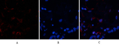
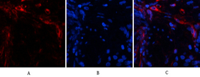
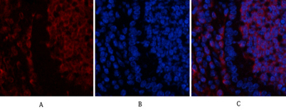
| Applications | IF;WB;IHC;ELISA |
| Other name | UBB; Polyubiquitin-B; UBC; Polyubiquitin-C; RPS27A; UBA80; UBCEP1; Ubiquitin-40S ribosomal protein S27a; Ubiquitin carboxyl extension protein 80; UBA52; UBCEP2; Ubiquitin-60S ribosomal protein L40; CEP52; Ubiquitin A-52 residue ribosomal protein fusion product 1 |
| Size | 50μL |
| Unit price ($) | 148 |
| Human gene ID | |
| Human Swiss-Prot | P62987/P62979/P0CG47/P0CG48 |
| Source | Rabbit |
| Isotype | IgG |
| Target | Ub |
| Fields | >>Ribosome;>>Ubiquitin mediated proteolysis;>>Mitophagy - animal;>>Parkinson disease;>>Pathways of neurodegeneration - multiple diseases;>>Shigellosis;>>Kaposi sarcoma-associated herpesvirus infection;>>Coronavirus disease - COVID-19 |
| Gene name | UBA52/RPS27A/UBB/UBC |
| Protein name | Ubiquitin |
| Human gene link | |
| Human Swiss link | View Human Swiss-Prot |
| Mouse gene ID | |
| Mouse gene link | |
| Mouse Swiss-Prot | |
| Mouse Swiss link | |
| Rat gene ID | |
| Rat gene link | |
| Rat Swiss-Prot | |
| Rat Swiss link | |
| Immunogen | Synthesized peptide derived from the N-terminal region of human Ub. |
| Specificity | Ub Polyclonal Antibody detects endogenous levels of Ub protein. |
| Formulation | Liquid in PBS containing 50% glycerol, 0.5% BSA and 0.02% sodium azide. |
| Clonality | Polyclonal |
| Dilution | IF: 1:50-200 Western Blot: 1/500 - 1/2000. IHC-p: 1:100-1:300. ELISA: 1/10000. Not yet tested in other applications. |
| Purification | The antibody was affinity-purified from rabbit antiserum by affinity-chromatography using epitope-specific immunogen. |
| Concentration | 1 mg/ml |
| Storage stability | -20°C/1 year |
| Molecular Weight (Da) | |
| Observed band (KD) | 80 50kD |
| Background | Ubiquitin is a highly conserved nuclear and cytoplasmic protein that has a major role in targeting cellular proteins for degradation by the 26S proteosome. It is also involved in the maintenance of chromatin structure, the regulation of gene expression, and the stress response. Ubiquitin is synthesized as a precursor protein consisting of either polyubiquitin chains or a single ubiquitin moiety fused to an unrelated protein. This gene encodes a fusion protein consisting of ubiquitin at the N terminus and ribosomal protein L40 at the C terminus, a C-terminal extension protein (CEP). Multiple processed pseudogenes derived from this gene are present in the genome. [provided by RefSeq, Jul 2008], |
| Function | function:Protein modifier which can be covalently attached to target lysines either as a monomer or as a lysine-linked polymer. Attachment to proteins as a Lys-48-linked polymer usually leads to their degradation by proteasome. Attachment to proteins as a monomer or as an alternatively linked polymer does not lead to proteasomal degradation and may be required for numerous functions, including maintenance of chromatin structure, regulation of gene expression, stress response, ribosome biogenesis and DNA repair.,miscellaneous:This ribosomal protein is synthesized as a C-terminal extension protein (CEP) of ubiquitin.,miscellaneous:Ubiquitin is synthesized as a polyubiquitin precursor with exact head to tail repeats, the number of repeats differ between species and strains. In some species there is a final amino-acid after the last repeat, here in human a Val. Some ubiquitin genes contain a |
| Subcellular location | [Ubiquitin]: Cytoplasm . Nucleus .; [60S ribosomal protein L40]: Cytoplasm . |
| Expression | Brain,Epithelium,Fetal brain cortex,Liver,L |

Use collapsible tabs for more detailed information that will help customers make a purchasing decision.
Ex: Shipping and return policies, size guides, and other common questions.