 Français
 English

| ELK.No | ES4279 |
| Product name | FGF-2 rabbit pAb |
| Reactivity | Human;Mouse;Rat |
| Applications | WB;IHC;IF;ELISA |
| Other name | FGF2; FGFB; Fibroblast growth factor 2; FGF-2; Basic fibroblast growth factor; bFGF; Heparin-binding growth factor 2; HBGF-2 |
| Size | 50μL |
| Unit price ($) | 148 |
| Human gene ID | 2247 |
| Human Swiss-Prot | P09038 |
| Source | Rabbit |
| Isotype | IgG |
| Target | FGF-2 |
| Fields | >>EGFR tyrosine kinase inhibitor resistance;>>MAPK signaling pathway;>>Ras signaling pathway;>>Rap1 signaling pathway;>>Calcium signaling pathway;>>PI3K-Akt signaling pathway;>>Signaling pathways regulating pluripotency of stem cells;>>Regulation of actin cytoskeleton;>>Kaposi sarcoma-associated herpesvirus infection;>>Pathways in cancer;>>Proteoglycans in cancer;>>Chemical carcinogenesis - receptor activation;>>Melanoma;>>Breast cancer;>>Gastric cancer |
| Gene name | FGF2 |
| Protein name | Fibroblast growth factor 2 |
| Human gene link | View Human Gene |
| Human Swiss link | View Human Swiss-Prot |
| Mouse gene ID | 14173 |
| Mouse gene link | View Mouse Gene |
| Mouse Swiss-Prot | P15655 |
| Mouse Swiss link | View Mouse Swiss-Prot |
| Rat gene ID | 54250 |
| Rat gene link | View Rat Gene |
| Rat Swiss-Prot | P13109 |
| Rat Swiss link | View Rat Swiss-Prot |
| Immunogen | The antiserum was produced against synthesized peptide derived from the Internal region of human FGF2. AA range:151-200 |
| Specificity | FGF-2 Polyclonal Antibody detects endogenous levels of FGF-2 protein. |
| Formulation | Liquid in PBS containing 50% glycerol, 0.5% BSA and 0.02% sodium azide. |
| Clonality | Polyclonal |
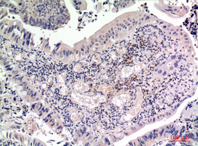
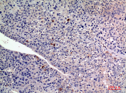
| Dilution | Western Blot: 1/500 - 1/2000. IHC-p: 1:100-1:300. ELISA: 1/10000. Not yet tested in other applications. |
| Purification | The antibody was affinity-purified from rabbit antiserum by affinity-chromatography using epitope-specific immunogen. |
| Concentration | 1 mg/ml |
| Storage stability | -20°C/1 year |
| Molecular Weight (Da) | |
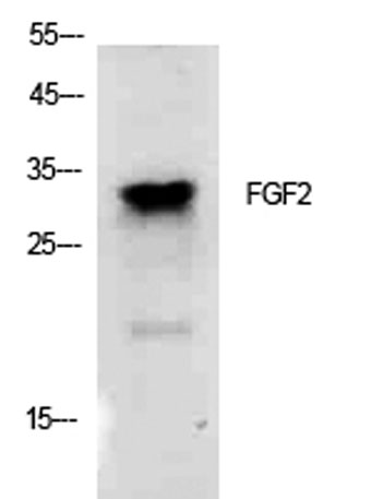
| Observed band (KD) | 30kD |
| Background | The protein encoded by this gene is a member of the fibroblast growth factor (FGF) family. FGF family members bind heparin and possess broad mitogenic and angiogenic activities. This protein has been implicated in diverse biological processes, such as limb and nervous system development, wound healing, and tumor growth. The mRNA for this gene contains multiple polyadenylation sites, and is alternatively translated from non-AUG (CUG) and AUG initiation codons, resulting in five different isoforms with distinct properties. The CUG-initiated isoforms are localized in the nucleus and are responsible for the intracrine effect, whereas, the AUG-initiated form is mostly cytosolic and is responsible for the paracrine and autocrine effects of this FGF. [provided by RefSeq, Jul 2008], |
| Function | function:The heparin-binding growth factors are angiogenic agents in vivo and are potent mitogens for a variety of cell types in vitro. There are differences in the tissue distribution and concentration of these 2 growth factors.,miscellaneous:This protein binds heparin more strongly than does aFGF.,PTM:Several N-termini starting at positions 48, 54, 47 and 65 have been identified by direct sequencing.,sequence caution:Unusual initiator. The initiator methionine is coded by a non-canonical CTG leucine codon.,similarity:Belongs to the heparin-binding growth factors family.,subunit:Monomer. Interacts with CSPG4 and FGFBP1. Found in a complex with FGFBP1, FGF1 and FGF2.,tissue specificity:Expressed in granulosa and cumulus cells., |
| Subcellular location | Secreted . Nucleus . Exported from cells by an endoplasmic reticulum (ER)/Golgi-independent mechanism. Unconventional secretion of FGF2 occurs by direct translocation across the plasma membrane (PubMed:20230531). Binding of exogenous FGF2 to FGFR facilitates endocytosis followed by translocation of FGF2 across endosomal membrane into the cytosol (PubMed:22321063). Nuclear import from the cytosol requires the classical nuclear import machinery, involving proteins KPNA1 and KPNB1, as well as CEP57 (PubMed:22321063). . |
| Expression | Expressed in granulosa and cumulus cells. Expressed in hepatocellular carcinoma cells, but not in non-cancerous liver tissue. |

Use collapsible tabs for more detailed information that will help customers make a purchasing decision.
Ex: Shipping and return policies, size guides, and other common questions.