 Français
 English
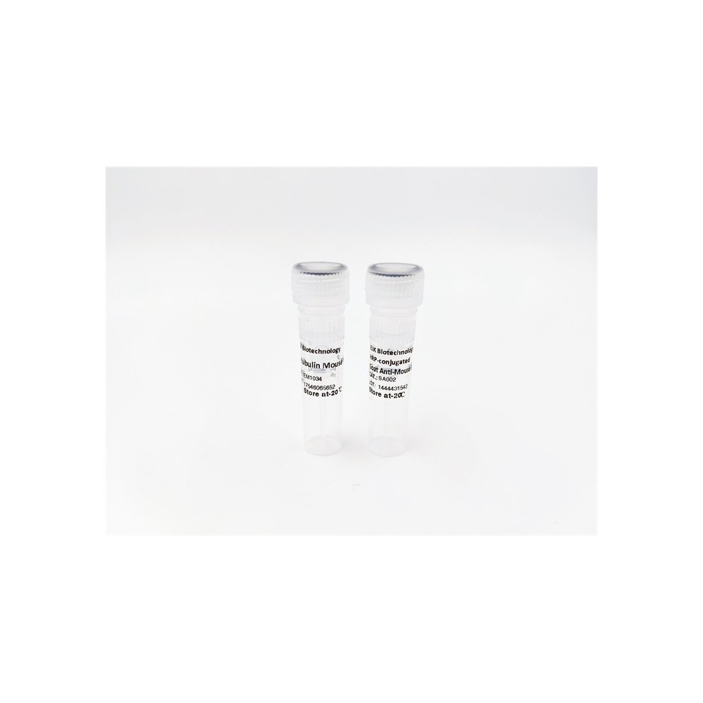

| ELK.No | ES4309 |
| Product name | CD13 rabbit pAb |
| Reactivity | Human;Rat;Mouse; |
| Applications | WB;IHC;IF;ELISA |
| Other name | ANPEP; APN; CD13; PEPN; Aminopeptidase N; AP-N; hAPN; Alanyl aminopeptidase; Aminopeptidase M; AP-M; Microsomal aminopeptidase; Myeloid plasma membrane glycoprotein CD13; gp150; CD13 |
| Size | 50μL |
| Unit price ($) | 148 |
| Human gene ID | 290 |
| Human Swiss-Prot | P15144 |
| Source | Rabbit |
| Isotype | IgG |
| Target | CD13 |
| Fields | >>Glutathione metabolism;>>Metabolic pathways;>>Renin-angiotensin system;>>Hematopoietic cell lineage |
| Gene name | ANPEP |
| Protein name | Aminopeptidase N |
| Human gene link | View Human Gene |
| Human Swiss link | View Human Swiss-Prot |
| Mouse gene ID | |
| Mouse gene link | |
| Mouse Swiss-Prot | P97449 |
| Mouse Swiss link | View Mouse Swiss-Prot |
| Rat gene ID | |
| Rat gene link | |
| Rat Swiss-Prot | |
| Rat Swiss link | |
| Immunogen | Synthesized peptide derived from Aminopeptidase N at AA range: 881-930 |
| Specificity | CD13 Polyclonal Antibody detects endogenous levels of CD13 protein. |
| Formulation | Liquid in PBS containing 50% glycerol, 0.5% BSA and 0.02% sodium azide. |
| Clonality | Polyclonal |
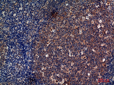
| Dilution | Western Blot: 1/500 - 1/2000. IHC-p: 1:100-1:300. ELISA: 1/10000. Not yet tested in other applications. |
| Purification | The antibody was affinity-purified from rabbit antiserum by affinity-chromatography using epitope-specific immunogen. |
| Concentration | 1 mg/ml |
| Storage stability | -20°C/1 year |
| Molecular Weight (Da) | |
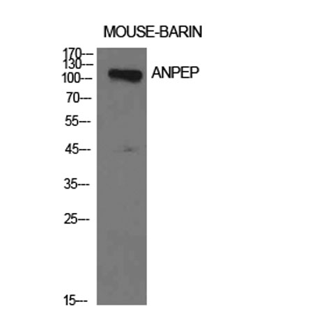
| Observed band (KD) | 110kD |
| Background | Aminopeptidase N is located in the small-intestinal and renal microvillar membrane, and also in other plasma membranes. In the small intestine aminopeptidase N plays a role in the final digestion of peptides generated from hydrolysis of proteins by gastric and pancreatic proteases. Its function in proximal tubular epithelial cells and other cell types is less clear. The large extracellular carboxyterminal domain contains a pentapeptide consensus sequence characteristic of members of the zinc-binding metalloproteinase superfamily. Sequence comparisons with known enzymes of this class showed that CD13 and aminopeptidase N are identical. The latter enzyme was thought to be involved in the metabolism of regulatory peptides by diverse cell types, including small intestinal and renal tubular epithelial cells, macrophages, granulocytes, and synaptic membranes from the CNS. Human aminopeptidase N is |
| Function | catalytic activity:Release of an N-terminal amino acid, Xaa-|-Yaa- from a peptide, amide or arylamide. Xaa is preferably Ala, but may be most amino acids including Pro (slow action). When a terminal hydrophobic residue is followed by a prolyl residue, the two may be released as an intact Xaa-Pro dipeptide.,cofactor:Binds 1 zinc ion per subunit.,disease:Defects in ANPEP may be a cause of various types of leukemia or lymphoma.,domain:Amino acids 260-353 are essential to mediate susceptibility to infection with HCoV-229E (in porcine/human chimeric studies) and more specifically amino acids 288-295 (mutagenesis studies).,function:Broad specificity aminopeptidase. Plays a role in the final digestion of peptides generated from hydrolysis of proteins by gastric and pancreatic proteases. May play a critical role in the pathogenesis of cholesterol gallstone disease. May be involved in the metabol |
| Subcellular location | Cell membrane ; Single-pass type II membrane protein . Also found as a soluble form. . |
| Expression | Expressed in epithelial cells of the kidney, intestine, and respiratory tract; granulocytes, monocytes, fibroblasts, endothelial cells, cerebral pericytes at the blood-brain barrier, synaptic membranes of cells in the CNS. Also expressed in endometrial stromal cells, but not in the endometrial glandular cells. Found in the vasculature of tissues that undergo angiogenesis and in malignant gliomas and lymph node metastases from multiple tumor types but not in blood vessels of normal tissues. A soluble form has been found in plasma. It is found to be elevated in plasma and effusions of cancer patients. |

Use collapsible tabs for more detailed information that will help customers make a purchasing decision.
Ex: Shipping and return policies, size guides, and other common questions.