 Français
 English
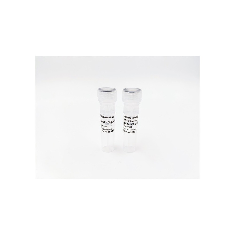

| ELK.No | ES4400 |
| Product name | Troponin I-C rabbit pAb |
| Reactivity | Mouse;Rat |
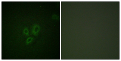
| Applications | WB;IHC;IF;ELISA |
| Other name | TNNI3; TNNC1; Troponin I; cardiac muscle; Cardiac troponin I |
| Size | 50μL |
| Unit price ($) | 148 |
| Human gene ID | |
| Human Swiss-Prot | |
| Source | Rabbit |
| Isotype | IgG |
| Target | Troponin I-C |
| Fields | |
| Gene name | TNNI3 |
| Protein name | Troponin I cardiac muscle |
| Human gene link | |
| Human Swiss link | |
| Mouse gene ID | 21954 |
| Mouse gene link | View Mouse Gene |
| Mouse Swiss-Prot | |
| Mouse Swiss link | |
| Rat gene ID | 29248 |
| Rat gene link | View Rat Gene |
| Rat Swiss-Prot | P23693 |
| Rat Swiss link | View Rat Swiss-Prot |
| Immunogen | The antiserum was produced against synthesized peptide derived from mouse TNNI3. AA range:5-54 |
| Specificity | Troponin I-C Polyclonal Antibody detects endogenous levels of Troponin I-C protein. |
| Formulation | Liquid in PBS containing 50% glycerol, 0.5% BSA and 0.02% sodium azide. |
| Clonality | Polyclonal |
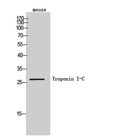
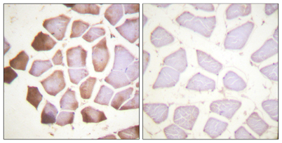
| Dilution | Western Blot: 1/500 - 1/2000. Immunohistochemistry: 1/100 - 1/300. ELISA: 1/5000. Not yet tested in other applications. |
| Purification | The antibody was affinity-purified from rabbit antiserum by affinity-chromatography using epitope-specific immunogen. |
| Concentration | 1 mg/ml |
| Storage stability | -20°C/1 year |
| Molecular Weight (Da) | |
| Observed band (KD) | 28kD |
| Background | Troponin I (TnI), along with troponin T (TnT) and troponin C (TnC), is one of 3 subunits that form the troponin complex of the thin filaments of striated muscle. TnI is the inhibitory subunit; blocking actin-myosin interactions and thereby mediating striated muscle relaxation. The TnI subfamily contains three genes: tnI-skeletal-fast-twitch, TnI-skeletal-slow-twitch, and TnI-cardiac. This gene encodes the TnI-cardiac protein and is exclusively expressed in cardiac muscle tissues. Mutations in this gene cause familial hypertrophic cardiomyopathy type 7 (CMH7) and familial restrictive cardiomyopathy (RCM). |
| Function | |
| Subcellular location | |
| Expression |

Use collapsible tabs for more detailed information that will help customers make a purchasing decision.
Ex: Shipping and return policies, size guides, and other common questions.