 Français
 English

| ELK.No | ES4419 |
| Product name | Cadherin-8 rabbit pAb |
| Reactivity | Human;Mouse;Rat |
| Applications | WB;IHC;IF;ELISA |
| Other name | CDH8; Cadherin-8 |
| Size | 100μL |
| Unit price ($) | 248 |
| Human gene ID | 1006 |
| Human Swiss-Prot | P55286 |
| Source | Rabbit |
| Isotype | IgG |
| Target | Cadherin-8 |
| Fields | |
| Gene name | CDH8 |
| Protein name | Cadherin-8 |
| Human gene link | View Human Gene |
| Human Swiss link | View Human Swiss-Prot |
| Mouse gene ID | 12564 |
| Mouse gene link | View Mouse Gene |
| Mouse Swiss-Prot | P97291 |
| Mouse Swiss link | View Mouse Swiss-Prot |
| Rat gene ID | |
| Rat gene link | |
| Rat Swiss-Prot | O54800 |
| Rat Swiss link | View Rat Swiss-Prot |
| Immunogen | The antiserum was produced against synthesized peptide derived from human CDH8. AA range:491-540 |
| Specificity | Cadherin-8 Polyclonal Antibody detects endogenous levels of Cadherin-8 protein. |
| Formulation | Liquid in PBS containing 50% glycerol, 0.5% BSA and 0.02% sodium azide. |
| Clonality | Polyclonal |
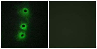
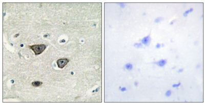
| Dilution | Western Blot: 1/500 - 1/2000. Immunohistochemistry: 1/100 - 1/300. Immunofluorescence: 1/200 - 1/1000. ELISA: 1/20000. Not yet tested in other applications. |
| Purification | The antibody was affinity-purified from rabbit antiserum by affinity-chromatography using epitope-specific immunogen. |
| Concentration | 1 mg/ml |
| Storage stability | -20°C/1 year |
| Molecular Weight (Da) | |
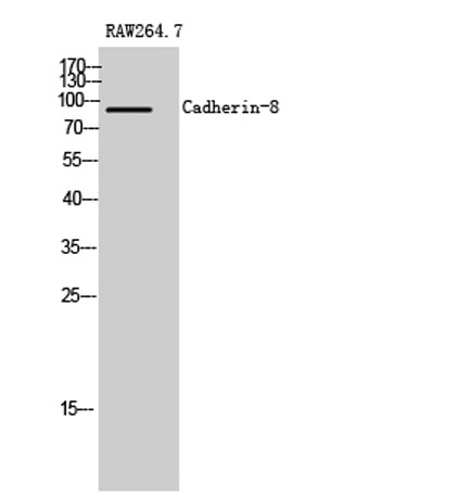
| Observed band (KD) | 90kD |
| Background | This gene encodes a type II classical cadherin from the cadherin superfamily, integral membrane proteins that mediate calcium-dependent cell-cell adhesion. Mature cadherin proteins are composed of a large N-terminal extracellular domain, a single membrane-spanning domain, and a small, highly conserved C-terminal cytoplasmic domain. The extracellular domain consists of 5 subdomains, each containing a cadherin motif, and appears to determine the specificity of the protein's homophilic cell adhesion activity. Type II (atypical) cadherins are defined based on their lack of a HAV cell adhesion recognition sequence specific to type I cadherins. This particular cadherin is expressed in brain and is putatively involved in synaptic adhesion, axon outgrowth and guidance. [provided by RefSeq, Jul 2008], |
| Function | function:Cadherins are calcium dependent cell adhesion proteins. They preferentially interact with themselves in a homophilic manner in connecting cells; cadherins may thus contribute to the sorting of heterogeneous cell types.,similarity:Contains 5 cadherin domains.,tissue specificity:Mainly expressed in brain. Found in certain nerve cell lines, such as retinoblasts, glioma cells and neuroblasts., |
| Subcellular location | Cell membrane; Single-pass type I membrane protein. |
| Expression | Mainly expressed in brain. Found in certain nerve cell lines, such as retinoblasts, glioma cells and neuroblasts. |

Use collapsible tabs for more detailed information that will help customers make a purchasing decision.
Ex: Shipping and return policies, size guides, and other common questions.