 Français
 English
ACK (phospho Tyr284) rabbit pAb
AO-06-ES4434-100

| ELK.No | ES4434 |
| Product name | ACK (phospho Tyr284) rabbit pAb |
| Reactivity | Human;Mouse |
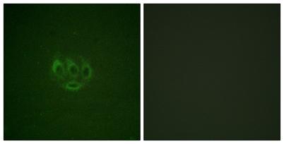
| Applications | WB;IHC;IF;ELISA |
| Other name | TNK2; ACK1; Activated CDC42 kinase 1; ACK-1; Tyrosine kinase non-receptor protein 2 |
| Size | 100μL |
| Unit price ($) | 248 |
| Human gene ID | 10188 |
| Human Swiss-Prot | Q07912 |
| Source | Rabbit |
| Isotype | IgG |
| Target | ACK |
| Fields | |
| Gene name | TNK2 |
| Protein name | Activated CDC42 kinase 1 |
| Human gene link | View Human Gene |
| Human Swiss link | View Human Swiss-Prot |
| Mouse gene ID | 51789 |
| Mouse gene link | View Mouse Gene |
| Mouse Swiss-Prot | O54967 |
| Mouse Swiss link | View Mouse Swiss-Prot |
| Rat gene ID | |
| Rat gene link | |
| Rat Swiss-Prot | |
| Rat Swiss link | |
| Immunogen | The antiserum was produced against synthesized peptide derived from human ACK1 around the phosphorylation site of Tyr284. AA range:250-299 |
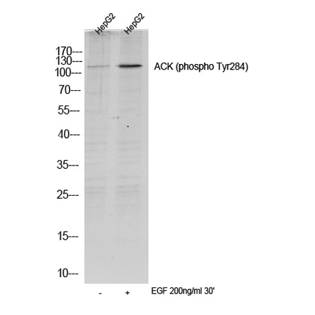
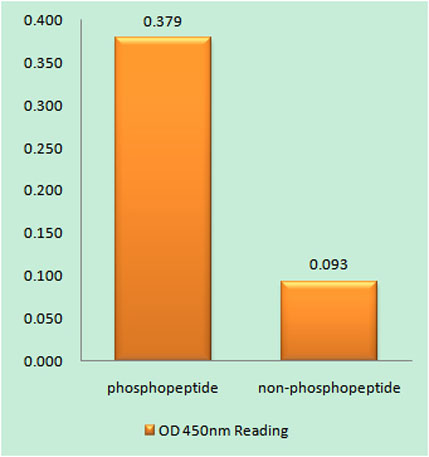
| Specificity | Phospho-ACK (Y284) Polyclonal Antibody detects endogenous levels of ACK protein only when phosphorylated at Y284. |
| Formulation | Liquid in PBS containing 50% glycerol, 0.5% BSA and 0.02% sodium azide. |
| Clonality | Polyclonal |
| Dilution | Western Blot: 1/500 - 1/2000. Immunohistochemistry: 1/100 - 1/300. Immunofluorescence: 1/200 - 1/1000. ELISA: 1/10000. Not yet tested in other applications. |
| Purification | The antibody was affinity-purified from rabbit antiserum by affinity-chromatography using epitope-specific immunogen. |
| Concentration | 1 mg/ml |
| Storage stability | -20°C/1 year |
| Molecular Weight (Da) | |
| Observed band (KD) | 120kD |
| Background | This gene encodes a tyrosine kinase that binds Cdc42Hs in its GTP-bound form and inhibits both the intrinsic and GTPase-activating protein (GAP)-stimulated GTPase activity of Cdc42Hs. This binding is mediated by a unique sequence of 47 amino acids C-terminal to an SH3 domain. The protein may be involved in a regulatory mechanism that sustains the GTP-bound active form of Cdc42Hs and which is directly linked to a tyrosine phosphorylation signal transduction pathway. Several alternatively spliced transcript variants have been identified from this gene, but the full-length nature of only two transcript variants has been determined. [provided by RefSeq, Jul 2008], |
| Function | catalytic activity:ATP + a [protein]-L-tyrosine = ADP + a [protein]-L-tyrosine phosphate.,cofactor:Magnesium.,enzyme regulation:The SH3 domain appears to play an autoinhibitory role.,function:Downstream effector of CDC42 which mediates CDC42-dependent cell migration via phosphorylation of BCAR1. Binds to both poly- and mono-ubiquitin and regulates ligand-induced degradation of EGFR. Participates in clathrin-mediated endocytosis. May be involved both in adult synaptic function and plasticity and in brain development.,sequence caution:Unlikely isoform. Aberrant splice sites.,similarity:Belongs to the protein kinase superfamily. Tyr protein kinase family.,similarity:Contains 1 CRIB domain.,similarity:Contains 1 protein kinase domain.,similarity:Contains 1 SH3 domain.,subunit:Interacts with CDC42. Interacts with activated CSPG4., |
| Subcellular location | Cell membrane . Nucleus . Endosome . Cell junction, adherens junction . Cytoplasmic vesicle membrane; Peripheral membrane protein; Cytoplasmic side . Cytoplasmic vesicle, clathrin-coated vesicle . Membrane, clathrin-coated pit . Cytoplasm, perinuclear region . Cytoplasm, cytosol . The Tyr-284 phosphorylated form is found both in the membrane and nucleus (By similarity). Co-localizes with EGFR on endosomes (PubMed:20333297). Nuclear translocation is CDC42-dependent (By similarity). Detected in long filamentous cytosolic structures where it co-localizes with CTPS1 (By similarity). . |
| Expression | The Tyr-284 phosphorylated form shows a significant increase in expression in breast cancers during the progressive stages i.e. normal to hyperplasia (ADH), ductal carcinoma in situ (DCIS), invasive ductal carcinoma (IDC) and lymph node metastatic (LNMM) stages. It also shows a significant increase in expression in prostate cancers during the progressive stages. |

Use collapsible tabs for more detailed information that will help customers make a purchasing decision.
Ex: Shipping and return policies, size guides, and other common questions.