 Français
 English

| ELK.No | ES4696 |
| Product name | Histone H4 rabbit pAb |
| Reactivity | Human;Mouse;Rat;Monkey |
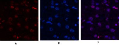
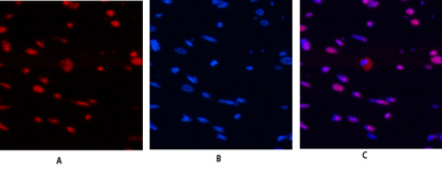
| Applications | WB;IHC;IF;ELISA |
| Other name | HIST1H4A; H4/A; H4FA; HIST1H4B; H4/I; H4FI; HIST1H4C; H4/G; H4FG; HIST1H4D; H4/B; H4FB; HIST1H4E; H4/J; H4FJ; HIST1H4F; H4/C; H4FC; HIST1H4H; H4/H; H4FH; HIST1H4I; H4/M; H4FM; HIST1H4J; H4/E; H4FE; HIST1H4K; H4/D; H4FD; HIST1H4L; H4/K; H4FK |
| Size | 50μL |
| Unit price ($) | 148 |
| Human gene ID | 121504/554313/8294/8359/8360/8361/8362/8363/8364/8365/8366/8367/8368/8370 |
| Human Swiss-Prot | P62805 |
| Source | Rabbit |
| Isotype | IgG |
| Target | Histone H4 |
| Fields | >>Neutrophil extracellular trap formation;>>Alcoholism;>>Viral carcinogenesis;>>Systemic lupus erythematosus |
| Gene name | HIST1H4A/HIST1H4B/HIST1H4C/HIST1H4D/HIST1H4E/HIST1H4F/HIST1H4H/HIST1H4I/HIST1H4J/HIST1H4K/HIST1H4L/HIST2H4A/HIST2H4B/HIST4H4 |
| Protein name | Histone H4 |
| Human gene link | View Human Gene |
| Human Swiss link | View Human Swiss-Prot |
| Mouse gene ID | |
| Mouse gene link | View Mouse Gene |
| Mouse Swiss-Prot | P62806 |
| Mouse Swiss link | View Mouse Swiss-Prot |
| Rat gene ID | 1.00361e+008 |
| Rat gene link | View Rat Gene |
| Rat Swiss-Prot | P62804 |
| Rat Swiss link | View Rat Swiss-Prot |
| Immunogen | The antiserum was produced against synthesized peptide derived from human Histone H4. AA range:6-55 |
| Specificity | Histone H4 Polyclonal Antibody detects endogenous levels of Histone H4 protein. |
| Formulation | Liquid in PBS containing 50% glycerol, 0.5% BSA and 0.02% sodium azide. |
| Clonality | Polyclonal |
| Dilution | Western Blot: 1/500 - 1/2000. Immunohistochemistry: 1/100 - 1/300. Immunofluorescence: 1/200 - 1/1000. ELISA: 1/5000. Not yet tested in other applications. |
| Purification | The antibody was affinity-purified from rabbit antiserum by affinity-chromatography using epitope-specific immunogen. |
| Concentration | 1 mg/ml |
| Storage stability | -20°C/1 year |
| Molecular Weight (Da) | |
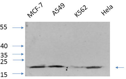
| Observed band (KD) | 11kD |
| Background | Histones are basic nuclear proteins that are responsible for the nucleosome structure of the chromosomal fiber in eukaryotes. Two molecules of each of the four core histones (H2A, H2B, H3, and H4) form an octamer, around which approximately 146 bp of DNA is wrapped in repeating units, called nucleosomes. The linker histone, H1, interacts with linker DNA between nucleosomes and functions in the compaction of chromatin into higher order structures. This gene is intronless and encodes a replication-dependent histone that is a member of the histone H4 family. Transcripts from this gene lack polyA tails but instead contain a palindromic termination element. This gene is found in the histone microcluster on chromosome 6p21.33. [provided by RefSeq, Aug 2015], |
| Function | function:Core component of nucleosome. Nucleosomes wrap and compact DNA into chromatin, limiting DNA accessibility to the cellular machineries which require DNA as a template. Histones thereby play a central role in transcription regulation, DNA repair, DNA replication and chromosomal stability. DNA accessibility is regulated via a complex set of post-translational modifications of histones, also called histone code, and nucleosome remodeling.,PTM:Acetylation at Lys-6, Lys-9, Lys-13 and Lys-17 occurs in coding regions of the genome but not in heterochromatin.,PTM:Citrullination at Arg-4 by PADI4 impairs methylation.,PTM:Monomethylated, dimethylated or trimethylated at Lys-21. Monomethylation is performed by SET8. Trimethylation is performed by SUV420H1 and SUV420H2 and induces gene silencing.,PTM:Monomethylation at Arg-4 by PRMT1 favors acetylation at Lys-9 and Lys-13. Demethylation is p |
| Subcellular location | Nucleus. Chromosome. |
| Expression | B-cell lymphoma,Bone marrow,Brain,Clones donated by HIP,Corpus call |

Use collapsible tabs for more detailed information that will help customers make a purchasing decision.
Ex: Shipping and return policies, size guides, and other common questions.