 Français
 English
PYK2 (phospho Tyr881) rabbit pAb
AO-06-ES5196-50

| ELK.No | ES5196 |
| Product name | PYK2 (phospho Tyr881) rabbit pAb |
| Reactivity | Human;Mouse;Rat |
| Applications | WB;IHC;IF;ELISA |
| Other name | PTK2B; FAK2; PYK2; RAFTK; Protein-tyrosine kinase 2-beta; Calcium-dependent tyrosine kinase; CADTK; Calcium-regulated non-receptor proline-rich tyrosine kinase; Cell adhesion kinase beta; CAK-beta; CAKB; Focal adhesion kinase 2; FADK 2; Pro |
| Size | 50μL |
| Unit price ($) | 148 |
| Human gene ID | 2185 |
| Human Swiss-Prot | Q14289 |
| Source | Rabbit |
| Isotype | IgG |
| Target | PYK2 |
| Fields | >>Calcium signaling pathway;>>Chemokine signaling pathway;>>Phospholipase D signaling pathway;>>Natural killer cell mediated cytotoxicity;>>Leukocyte transendothelial migration;>>GnRH signaling pathway;>>Yersinia infection;>>Hepatitis B;>>Human cytomegalovirus infection;>>Human immunodeficiency virus 1 infection |
| Gene name | PTK2B |
| Protein name | Protein-tyrosine kinase 2-beta |
| Human gene link | View Human Gene |
| Human Swiss link | View Human Swiss-Prot |
| Mouse gene ID | 19229 |
| Mouse gene link | View Mouse Gene |
| Mouse Swiss-Prot | Q9QVP9 |
| Mouse Swiss link | View Mouse Swiss-Prot |
| Rat gene ID | 50646 |
| Rat gene link | View Rat Gene |
| Rat Swiss-Prot | P70600 |
| Rat Swiss link | View Rat Swiss-Prot |
| Immunogen | The antiserum was produced against synthesized peptide derived from human PYK2 around the phosphorylation site of Tyr881. AA range:847-896 |
| Specificity | Phospho-PYK2 (Y881) Polyclonal Antibody detects endogenous levels of PYK2 protein only when phosphorylated at Y881. |
| Formulation | Liquid in PBS containing 50% glycerol, 0.5% BSA and 0.02% sodium azide. |
| Clonality | Polyclonal |
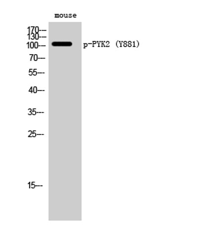
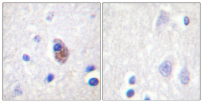
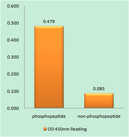
| Dilution | Western Blot: 1/500 - 1/2000. Immunohistochemistry: 1/100 - 1/300. ELISA: 1/5000. Not yet tested in other applications. |
| Purification | The antibody was affinity-purified from rabbit antiserum by affinity-chromatography using epitope-specific immunogen. |
| Concentration | 1 mg/ml |
| Storage stability | -20°C/1 year |
| Molecular Weight (Da) | |
| Observed band (KD) | 116kD |
| Background | This gene encodes a cytoplasmic protein tyrosine kinase which is involved in calcium-induced regulation of ion channels and activation of the map kinase signaling pathway. The encoded protein may represent an important signaling intermediate between neuropeptide-activated receptors or neurotransmitters that increase calcium flux and the downstream signals that regulate neuronal activity. The encoded protein undergoes rapid tyrosine phosphorylation and activation in response to increases in the intracellular calcium concentration, nicotinic acetylcholine receptor activation, membrane depolarization, or protein kinase C activation. This protein has been shown to bind CRK-associated substrate, nephrocystin, GTPase regulator associated with FAK, and the SH2 domain of GRB2. The encoded protein is a member of the FAK subfamily of protein tyrosine kinases but lacks significant sequence similarity t |
| Function | catalytic activity:ATP + a [protein]-L-tyrosine = ADP + a [protein]-L-tyrosine phosphate.,function:Involved in calcium induced regulation of ion channel and activation of the map kinase signaling pathway. May represent an important signaling intermediate between neuropeptide activated receptors or neurotransmitters that increase calcium flux and the downstream signals that regulate neuronal activity. Interacts with the SH2 domain of Grb2. May phosphorylate the voltage-gated potassium channel protein Kv1.2. Its activation is highly correlated with the stimulation of c-Jun N-terminal kinase activity. Involved in osmotic stress-dependent SNCA 'Tyr-125' phosphorylation.,PTM:Phosphorylated on tyrosine residues in response to various stimuli that elevate the intracellular calcium concentration, as well as by PKC activation. Recruitment by nephrocystin to cell matrix adhesions initiates Tyr-402 |
| Subcellular location | Cytoplasm. Cytoplasm, perinuclear region. Cell membrane; Peripheral membrane protein; Cytoplasmic side. Cell junction, focal adhesion. Cell projection, lamellipodium. Cytoplasm, cell cortex. Nucleus. Interaction with NPHP1 induces the membrane-association of the kinase. Colocalizes with integrins at the cell periphery. |
| Expression | Most abundant in the brain, with highest levels in amygdala and hippocampus. Low levels in kidney (at protein level). Also expressed in spleen and lymphocytes. |

Use collapsible tabs for more detailed information that will help customers make a purchasing decision.
Ex: Shipping and return policies, size guides, and other common questions.