 Français
 English
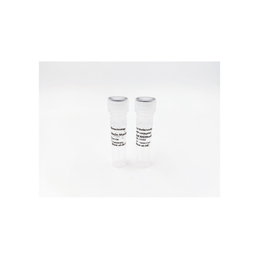

| ELK.No | ES5256 |
| Product name | TDAG51 rabbit pAb |
| Reactivity | Human;Mouse;Rat |
| Applications | WB;ELISA |
| Other name | PHLDA1; PHRIP; TDAG51; Pleckstrin homology-like domain family A member 1; Apoptosis-associated nuclear protein; Proline- and glutamine-rich protein; PQ-rich protein; PQR protein; Proline- and histidine-rich protein; T-cell death-associated |
| Size | 100μL |
| Unit price ($) | 248 |
| Human gene ID | 22822 |
| Human Swiss-Prot | Q8WV24 |
| Source | Rabbit |
| Isotype | IgG |
| Target | TDAG51 |
| Fields | |
| Gene name | PHLDA1 |
| Protein name | Pleckstrin homology-like domain family A member 1 |
| Human gene link | View Human Gene |
| Human Swiss link | View Human Swiss-Prot |
| Mouse gene ID | 21664 |
| Mouse gene link | View Mouse Gene |
| Mouse Swiss-Prot | Q62392 |
| Mouse Swiss link | View Mouse Swiss-Prot |
| Rat gene ID | |
| Rat gene link | |
| Rat Swiss-Prot | Q9QZA1 |
| Rat Swiss link | View Rat Swiss-Prot |
| Immunogen | The antiserum was produced against synthesized peptide derived from human PHLA1. AA range:271-320 |
| Specificity | TDAG51 Polyclonal Antibody detects endogenous levels of TDAG51 protein. |
| Formulation | Liquid in PBS containing 50% glycerol, 0.5% BSA and 0.02% sodium azide. |
| Clonality | Polyclonal |
| Dilution | Western Blot: 1/500 - 1/2000. ELISA: 1/10000. Not yet tested in other applications. |
| Purification | The antibody was affinity-purified from rabbit antiserum by affinity-chromatography using epitope-specific immunogen. |
| Concentration | 1 mg/ml |
| Storage stability | -20°C/1 year |
| Molecular Weight (Da) | |
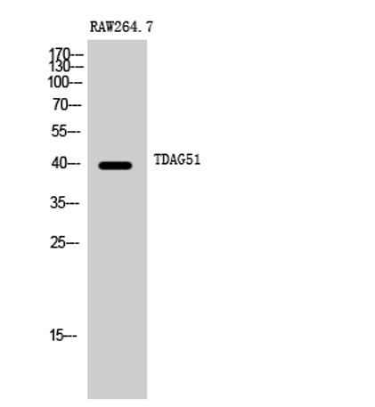
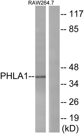
| Observed band (KD) | 40kD |
| Background | This gene encodes an evolutionarily conserved proline-histidine rich nuclear protein. The encoded protein may play an important role in the anti-apoptotic effects of insulin-like growth factor-1. [provided by RefSeq, Jul 2008], |
| Function | function:Seems to be involved in regulation of apoptosis. May be involved in detachment-mediated programmed cell death. May mediate apoptosis during neuronal development. May be involved in regulation of anti-apoptotic effects of IGF1. May be involved in translational regulation.,induction:Induced by homocysteine and other ER stress-inducing reagents. Induced by phorbolester (TPA)/ionomycin, and stimulation of the T-cell receptor (TCR) complex in T-cells.,similarity:Contains 1 PH domain.,subcellular location:Colocalizes with intracellular vesicles.,subunit:Interacts with RPL14, EIF3S7 and PABPC4.,tissue specificity:Widely expressed with highest levels in pancreas. Strongly expressed by benign melanocytic nevi, and progressively reduced expressed in primary and metastatic melanomas (at protein level)., |
| Subcellular location | Cytoplasm . Cytoplasmic vesicle . Nucleus, nucleolus . Colocalizes with intracellular vesicles. . |
| Expression | Widely expressed with highest levels in pancreas. Strongly expressed by benign melanocytic nevi, and progressively reduced expressed in primary and metastatic melanomas (at protein level). |

Use collapsible tabs for more detailed information that will help customers make a purchasing decision.
Ex: Shipping and return policies, size guides, and other common questions.