 Français
 English
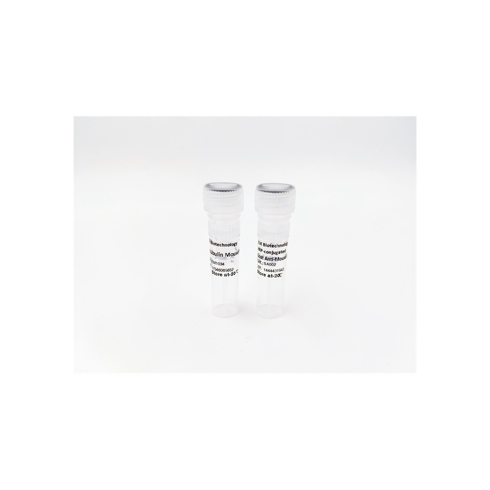

| ELK.No | ES5277 |
| Product name | Shank 2 rabbit pAb |
| Reactivity | Human;Rat;Mouse; |
| Applications | WB;IHC |
| Other name | SHANK2; CORTBP1; KIAA1022; SH3 and multiple ankyrin repeat domains protein 2; Shank2; Cortactin-binding protein 1; CortBP1; Proline-rich synapse-associated protein 1 |
| Size | 100μL |
| Unit price ($) | 248 |
| Human gene ID | 22941 |
| Human Swiss-Prot | Q9UPX8 |
| Source | Rabbit |
| Isotype | IgG |
| Target | Shank 2 |
| Fields | >>Glutamatergic synapse |
| Gene name | SHANK2 |
| Protein name | SH3 and multiple ankyrin repeat domains protein 2 |
| Human gene link | View Human Gene |
| Human Swiss link | View Human Swiss-Prot |
| Mouse gene ID | |
| Mouse gene link | |
| Mouse Swiss-Prot | Q80Z38 |
| Mouse Swiss link | View Mouse Swiss-Prot |
| Rat gene ID | |
| Rat gene link | |
| Rat Swiss-Prot | |
| Rat Swiss link | |
| Immunogen | The antiserum was produced against synthesized peptide derived from human SHANK2. AA range:331-380 |
| Specificity | Shank 2 Polyclonal Antibody detects endogenous levels of Shank 2 protein. |
| Formulation | Liquid in PBS containing 50% glycerol, 0.5% BSA and 0.02% sodium azide. |
| Clonality | Polyclonal |
| Dilution | WB 1:500-2000;IHC-p 1:50-300 |
| Purification | The antibody was affinity-purified from rabbit antiserum by affinity-chromatography using epitope-specific immunogen. |
| Concentration | 1 mg/ml |
| Storage stability | -20°C/1 year |
| Molecular Weight (Da) | |
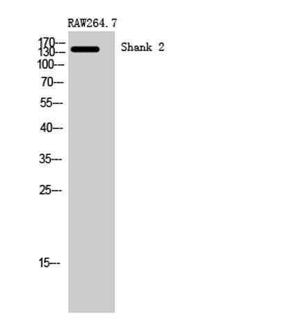
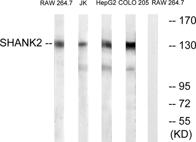
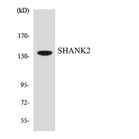
| Observed band (KD) | 135kD |
| Background | This gene encodes a protein that is a member of the Shank family of synaptic proteins that may function as molecular scaffolds in the postsynaptic density of excitatory synapses. Shank proteins contain multiple domains for protein-protein interaction, including ankyrin repeats, and an SH3 domain. This particular family member contains a PDZ domain, a consensus sequence for cortactin SH3 domain-binding peptides and a sterile alpha motif. The alternative splicing demonstrated in Shank genes has been suggested as a mechanism for regulating the molecular structure of Shank and the spectrum of Shank-interacting proteins in the postsynaptic densities of the adult and developing brain. Alterations in the encoded protein may be associated with susceptibility to autism spectrum disorder. Alternative splicing results in multiple transcript variants. [provided by RefSeq, Feb 2014], |
| Function | alternative products:Additional isoforms seem to exist,domain:The PDZ domain is required for interaction with GRID2, PLCB3, CFTR and SLC9A3.,function:Seems to be an adapter protein in the postsynaptic density (PSD) of excitatory synapses that interconnects receptors of the postsynaptic membrane including NMDA-type and metabotropic glutamate receptors, and the actin-based cytoskeleton. May play a role in the structural and functional organization of the dendritic spine and synaptic junction.,similarity:Belongs to the SHANK family.,similarity:Contains 1 PDZ (DHR) domain.,similarity:Contains 1 SAM (sterile alpha motif) domain.,subcellular location:Cytoplasm, postsynaptic density of neuronal cells.,subunit:Interacts with CCTN/cortactin SH3 domain, DLGAP1/GKAP and alpha-latrotoxin receptor 1. Is part of a complex with DLG4/PSD-95 and DLGAP1/GKAP. Interacts with GRID2, SLC9A3, CFTR and PLCB3. |
| Subcellular location | Apical cell membrane . Cytoplasm . Cell junction, synapse . Cell junction, synapse, postsynaptic density . Cell projection, growth cone . Cell projection, dendritic spine . Colocalizes with cortactin in growth cones in differentiating hippocampal neurons. Colocalized with PDE4D to the apical membrane of colonic crypt cells (By similarity). . |
| Expression | Isoform 3 is present in epithelial colonic cells (at protein level). |

Use collapsible tabs for more detailed information that will help customers make a purchasing decision.
Ex: Shipping and return policies, size guides, and other common questions.