 Français
 English

| ELK.No | ES5427 |
| Product name | GABAB R1 rabbit pAb |
| Reactivity | Human;Mouse;Rat;Guinea pig |
| Applications | WB;IF;ELISA |
| Other name | GABBR1; GPRC3A; Gamma-aminobutyric acid type B receptor subunit 1; GABA-B receptor 1; GABA-B-R1; GABA-BR1; GABABR1; Gb1 |
| Size | 100μL |
| Unit price ($) | 248 |
| Human gene ID | 2550 |
| Human Swiss-Prot | Q9UBS5 |
| Source | Rabbit |
| Isotype | IgG |
| Target | GABAB R1 |
| Fields | >>cAMP signaling pathway;>>Neuroactive ligand-receptor interaction;>>GABAergic synapse;>>Taste transduction;>>Estrogen signaling pathway;>>GnRH secretion;>>Morphine addiction |
| Gene name | GABBR1 |
| Protein name | Gamma-aminobutyric acid type B receptor subunit 1 |
| Human gene link | View Human Gene |
| Human Swiss link | View Human Swiss-Prot |
| Mouse gene ID | 54393 |
| Mouse gene link | View Mouse Gene |
| Mouse Swiss-Prot | Q9WV18 |
| Mouse Swiss link | View Mouse Swiss-Prot |
| Rat gene ID | 81657 |
| Rat gene link | View Rat Gene |
| Rat Swiss-Prot | Q9Z0U4 |
| Rat Swiss link | View Rat Swiss-Prot |
| Immunogen | The antiserum was produced against synthesized peptide derived from human GABBR1. AA range:891-940 |
| Specificity | GABAB R1 Polyclonal Antibody detects endogenous levels of GABAB R1 protein. |
| Formulation | Liquid in PBS containing 50% glycerol, 0.5% BSA and 0.02% sodium azide. |
| Clonality | Polyclonal |
| Dilution | Western Blot: 1/500 - 1/2000. Immunofluorescence: 1/200 - 1/1000. ELISA: 1/5000. Not yet tested in other applications. |
| Purification | The antibody was affinity-purified from rabbit antiserum by affinity-chromatography using epitope-specific immunogen. |
| Concentration | 1 mg/ml |
| Storage stability | -20°C/1 year |
| Molecular Weight (Da) | |
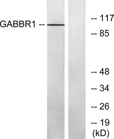
| Observed band (KD) | 108kD |
| Background | This gene encodes a receptor for gamma-aminobutyric acid (GABA), which is the main inhibitory neurotransmitter in the mammalian central nervous system. This receptor functions as a heterodimer with GABA(B) receptor 2. Defects in this gene may underlie brain disorders such as schizophrenia and epilepsy. Alternative splicing generates multiple transcript variants, but the full-length nature of some of these variants has not been determined. [provided by RefSeq, Jan 2016], |
| Function | alternative products:Isoforms corresponding to the full receptor are essentially found in the central nervous system (CNS),cofactor:Calcium. Required for high affinity binding to GABA.,domain:Alpha-helical parts of the C-terminal intracellular region mediate heterodimeric interaction with GABA-B receptor 2. The linker region between the transmembrane domain 3 (TM3) and the transmembrane domain 4 (TM4) probably play a role in the specificity for G-protein coupling.,function:Isoform 1E function may be to regulate the availability of functional GABA-B-R1A/GABA-B-R2 heterodimers by competing for GABA-B-R2 dimerization. This could explain the observation that certain small molecule ligands exhibit differential affinity for central versus peripheral sites.,function:Receptor for GABA. The activity of this receptor is mediated by G-proteins that inhibit adenylyl cyclase activity, stimulates phos |
| Subcellular location | Cell membrane ; Multi-pass membrane protein . Cell junction, synapse, postsynaptic cell membrane ; Multi-pass membrane protein . Cell projection, dendrite . Colocalizes with ATF4 in hippocampal neuron dendritic membranes (By similarity). Coexpression of GABBR1 and GABBR2 is required for GABBR1 maturation and transport to the plasma membrane (PubMed:15617512). .; [Isoform 1E]: Secreted . |
| Expression | Highly expressed in brain (PubMed:9844003, PubMed:9753614, PubMed:9872744). Weakly expressed in heart, small intestine and uterus. Isoform 1A: Mainly expressed in granular cell and molecular layer (PubMed:9844003). Isoform 1B: Mainly expressed in Purkinje cells (PubMed:9844003). Isoform 1E: Predominantly expressed in peripheral tissues as kidney, lung, trachea, colon, small intestine, stomach, bone marrow, thymus and mammary gland (PubMed:10906333). |


Use collapsible tabs for more detailed information that will help customers make a purchasing decision.
Ex: Shipping and return policies, size guides, and other common questions.