 Français
 English
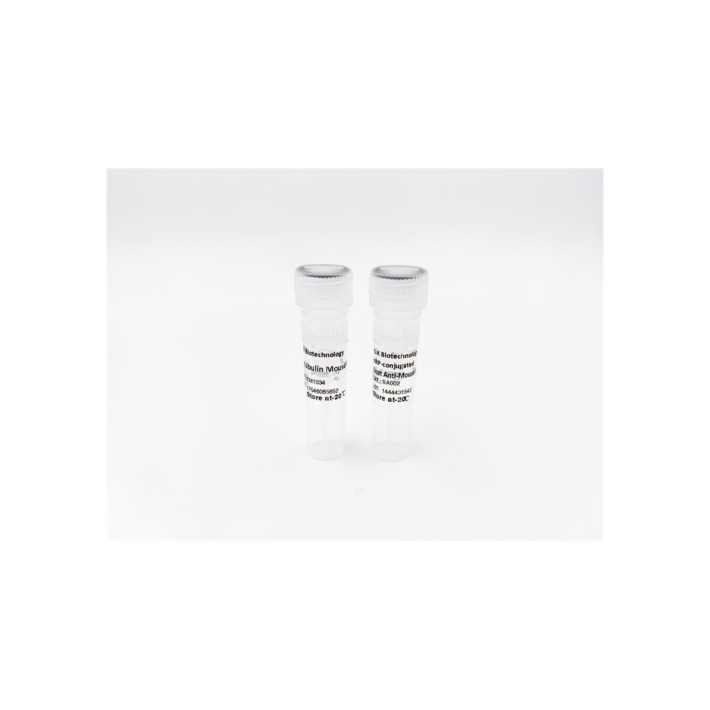

| ELK.No | ES5514 |
| Product name | p52 S6 kinase rabbit pAb |
| Reactivity | Human;Mouse |
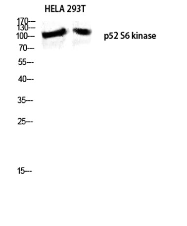
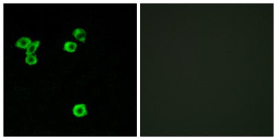
| Applications | WB;IF;ELISA |
| Other name | RPS6KC1; RPK118; Ribosomal protein S6 kinase delta-1; S6K-delta-1; 52 kDa ribosomal protein S6 kinase; Ribosomal S6 kinase-like protein with two PSK domains 118 kDa protein; SPHK1-binding protein |
| Size | 100μL |
| Unit price ($) | 248 |
| Human gene ID | 26750 |
| Human Swiss-Prot | Q96S38 |
| Source | Rabbit |
| Isotype | IgG |
| Target | p52 S6 kinase |
| Fields | |
| Gene name | RPS6KC1 |
| Protein name | Ribosomal protein S6 kinase delta-1 |
| Human gene link | View Human Gene |
| Human Swiss link | View Human Swiss-Prot |
| Mouse gene ID | 320119 |
| Mouse gene link | View Mouse Gene |
| Mouse Swiss-Prot | Q8BLK9 |
| Mouse Swiss link | View Mouse Swiss-Prot |
| Rat gene ID | |
| Rat gene link | |
| Rat Swiss-Prot | |
| Rat Swiss link | |
| Immunogen | The antiserum was produced against synthesized peptide derived from human RPS6KC1. AA range:231-280 |
| Specificity | p52 S6 kinase Polyclonal Antibody detects endogenous levels of p52 S6 kinase protein. |
| Formulation | Liquid in PBS containing 50% glycerol, 0.5% BSA and 0.02% sodium azide. |
| Clonality | Polyclonal |
| Dilution | Western Blot: 1/500 - 1/2000. Immunofluorescence: 1/200 - 1/1000. ELISA: 1/20000. Not yet tested in other applications. |
| Purification | The antibody was affinity-purified from rabbit antiserum by affinity-chromatography using epitope-specific immunogen. |
| Concentration | 1 mg/ml |
| Storage stability | -20°C/1 year |
| Molecular Weight (Da) | |
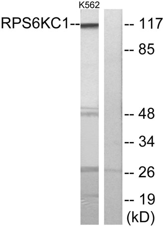
| Observed band (KD) | 117kD |
| Background | catalytic activity:ATP + a protein = ADP + a phosphoprotein.,caution:Instead of Lys-820, Arg-820 is found at the binding site.,domain:The first protein kinase domain appears to be a pseudokinase domain as it does not contain the classical characteristics, such as the ATP-binding motif, ATP-binding site and active site.,function:May be involved in transmitting sphingosine-1 phosphate (SPP)-mediated signaling into the cell.,similarity:Belongs to the protein kinase superfamily. Ser/Thr protein kinase family. S6 kinase subfamily.,similarity:Contains 1 MIT domain.,similarity:Contains 1 PX (phox homology) domain.,similarity:Contains 2 protein kinase domains.,subcellular location:Also found in some small dot-like or ring-shaped early endosome structures.,subunit:Interacts with SPHK1 and phosphatidylinositol 3-phosphate.,tissue specificity:Highly expressed in testis, skeletal muscle, brain, heart, placenta, kidney and liver and weakly expressed in thymus, small intestine, lung and colon., |
| Function | catalytic activity:ATP + a protein = ADP + a phosphoprotein.,caution:Instead of Lys-820, Arg-820 is found at the binding site.,domain:The first protein kinase domain appears to be a pseudokinase domain as it does not contain the classical characteristics, such as the ATP-binding motif, ATP-binding site and active site.,function:May be involved in transmitting sphingosine-1 phosphate (SPP)-mediated signaling into the cell.,similarity:Belongs to the protein kinase superfamily. Ser/Thr protein kinase family. S6 kinase subfamily.,similarity:Contains 1 MIT domain.,similarity:Contains 1 PX (phox homology) domain.,similarity:Contains 2 protein kinase domains.,subcellular location:Also found in some small dot-like or ring-shaped early endosome structures.,subunit:Interacts with SPHK1 and phosphatidylinositol 3-phosphate.,tissue specificity:Highly expressed in testis, skeletal muscle, brain, hear |
| Subcellular location | Cytoplasm . Membrane . Early endosome . |
| Expression | Highly expressed in testis, skeletal muscle, brain, heart, placenta, kidney and liver and weakly expressed in thymus, small intestine, lung and colon. |

Use collapsible tabs for more detailed information that will help customers make a purchasing decision.
Ex: Shipping and return policies, size guides, and other common questions.