 Français
 English
Lck BP-1 (phospho Tyr397) rabbit pAb
AO-06-ES5723-100

| ELK.No | ES5723 |
| Product name | Lck BP-1 (phospho Tyr397) rabbit pAb |
| Reactivity | Human;Rat;Mouse; |
| Applications | WB;IHC |
| Other name | HCLS1; HS1; Hematopoietic lineage cell-specific protein; Hematopoietic cell-specific LYN substrate 1; LckBP1; p75 |
| Size | 100μL |
| Unit price ($) | 248 |
| Human gene ID | 3059 |
| Human Swiss-Prot | P14317 |
| Source | Rabbit |
| Isotype | IgG |
| Target | Lck BP-1 |
| Fields | >>Tight junction;>>Bacterial invasion of epithelial cells;>>Pathogenic Escherichia coli infection;>>Shigellosis;>>Proteoglycans in cancer |
| Gene name | HCLS1 |
| Protein name | Hematopoietic lineage cell-specific protein |
| Human gene link | View Human Gene |
| Human Swiss link | View Human Swiss-Prot |
| Mouse gene ID | |
| Mouse gene link | |
| Mouse Swiss-Prot | P49710 |
| Mouse Swiss link | View Mouse Swiss-Prot |
| Rat gene ID | |
| Rat gene link | |
| Rat Swiss-Prot | |
| Rat Swiss link | |
| Immunogen | The antiserum was produced against synthesized peptide derived from human HS1 around the phosphorylation site of Tyr397. AA range:366-415 |
| Specificity | Phospho-Lck BP-1 (Y397) Polyclonal Antibody detects endogenous levels of Lck BP-1 protein only when phosphorylated at Y397. |
| Formulation | Liquid in PBS containing 50% glycerol, 0.5% BSA and 0.02% sodium azide. |
| Clonality | Polyclonal |
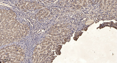
| Dilution | WB 1:500-2000;IHC-p 1:50-300 |
| Purification | The antibody was affinity-purified from rabbit antiserum by affinity-chromatography using epitope-specific immunogen. |
| Concentration | 1 mg/ml |
| Storage stability | -20°C/1 year |
| Molecular Weight (Da) | |
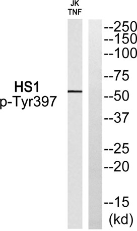
| Observed band (KD) | 55kD |
| Background | developmental stage:Expressed in early stage of myeloid and erythroid differentiation.,function:Substrate of the antigen receptor-coupled tyrosine kinase. Plays a role in antigen receptor signaling for both clonal expansion and deletion in lymphoid cells. May also be involved in the regulation of gene expression.,PTM:Phosphorylated by LYN; rapidly after cross-linking of surface IgM on B-cells.,similarity:Contains 1 SH3 domain.,similarity:Contains 4 cortactin repeats.,subunit:Associates with the SH2 and SH3 domains of LCK. Binding to he LCK SH3 domain occurs constitutively, while binding to the LCK SH2 domain occurs only upon TCR stimulation. A similar binding pattern was observed with LYN, but not with FYN in which the FYN SH2 region associates upon TCR stimulation but the FYN SH3 region does not associate regardless of TCR stimulation. Directly associates with HAX1, through binding to its C-terminal region. Interacts with HS1BP3.,tissue specificity:Expressed only in tissues and cells of hematopoietic origin., |
| Function | developmental stage:Expressed in early stage of myeloid and erythroid differentiation.,function:Substrate of the antigen receptor-coupled tyrosine kinase. Plays a role in antigen receptor signaling for both clonal expansion and deletion in lymphoid cells. May also be involved in the regulation of gene expression.,PTM:Phosphorylated by LYN; rapidly after cross-linking of surface IgM on B-cells.,similarity:Contains 1 SH3 domain.,similarity:Contains 4 cortactin repeats.,subunit:Associates with the SH2 and SH3 domains of LCK. Binding to he LCK SH3 domain occurs constitutively, while binding to the LCK SH2 domain occurs only upon TCR stimulation. A similar binding pattern was observed with LYN, but not with FYN in which the FYN SH2 region associates upon TCR stimulation but the FYN SH3 region does not associate regardless of TCR stimulation. Directly associates with HAX1, through binding to i |
| Subcellular location | Membrane ; Peripheral membrane protein . Cytoplasm . Mitochondrion . |
| Expression | Expressed only in tissues and cells of hematopoietic origin. |

Use collapsible tabs for more detailed information that will help customers make a purchasing decision.
Ex: Shipping and return policies, size guides, and other common questions.