 Français
 English

| ELK.No | ES5883 |
| Product name | CD130 rabbit pAb |
| Reactivity | Human;Mouse |
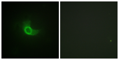
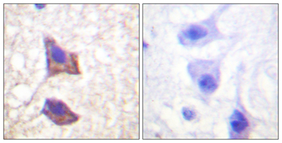
| Applications | WB;IHC;IF;ELISA |
| Other name | IL6ST; Interleukin-6 receptor subunit beta; IL-6 receptor subunit beta; IL-6R subunit beta; IL-6R-beta; IL-6RB; CDw130; Interleukin-6 signal transducer; Membrane glycoprotein 130; gp130; Oncostatin-M receptor subunit alpha; CD antigen CD130 |
| Size | 100μL |
| Unit price ($) | 248 |
| Human gene ID | 3572 |
| Human Swiss-Prot | P40189 |
| Source | Rabbit |
| Isotype | IgG |
| Target | CD130/gp130 |
| Fields | >>Cytokine-cytokine receptor interaction;>>Viral protein interaction with cytokine and cytokine receptor;>>Signaling pathways regulating pluripotency of stem cells;>>JAK-STAT signaling pathway;>>Th17 cell differentiation;>>Kaposi sarcoma-associated herpesvirus infection;>>Coronavirus disease - COVID-19;>>Pathways in cancer;>>Viral carcinogenesis |
| Gene name | IL6ST |
| Protein name | Interleukin-6 receptor subunit beta |
| Human gene link | View Human Gene |
| Human Swiss link | View Human Swiss-Prot |
| Mouse gene ID | 16195 |
| Mouse gene link | View Mouse Gene |
| Mouse Swiss-Prot | Q00560 |
| Mouse Swiss link | View Mouse Swiss-Prot |
| Rat gene ID | |
| Rat gene link | |
| Rat Swiss-Prot | |
| Rat Swiss link | |
| Immunogen | The antiserum was produced against synthesized peptide derived from human CD130/gp130. AA range:748-797 |
| Specificity | CD130 Polyclonal Antibody detects endogenous levels of CD130 protein. |
| Formulation | Liquid in PBS containing 50% glycerol, 0.5% BSA and 0.02% sodium azide. |
| Clonality | Polyclonal |
| Dilution | Western Blot: 1/500 - 1/2000. Immunohistochemistry: 1/100 - 1/300. Immunofluorescence: 1/200 - 1/1000. ELISA: 1/20000. Not yet tested in other applications. |
| Purification | The antibody was affinity-purified from rabbit antiserum by affinity-chromatography using epitope-specific immunogen. |
| Concentration | 1 mg/ml |
| Storage stability | -20°C/1 year |
| Molecular Weight (Da) | |
| Observed band (KD) | 160kD |
| Background | The protein encoded by this gene is a signal transducer shared by many cytokines, including interleukin 6 (IL6), ciliary neurotrophic factor (CNTF), leukemia inhibitory factor (LIF), and oncostatin M (OSM). This protein functions as a part of the cytokine receptor complex. The activation of this protein is dependent upon the binding of cytokines to their receptors. vIL6, a protein related to IL6 and encoded by the Kaposi sarcoma-associated herpesvirus, can bypass the interleukin 6 receptor (IL6R) and directly activate this protein. Knockout studies in mice suggest that this gene plays a critical role in regulating myocyte apoptosis. Alternatively spliced transcript variants have been described. A related pseudogene has been identified on chromosome 17. [provided by RefSeq, May 2014], |
| Function | disease:Isoform 2 is an autoantigen found in rheumatoid arthritis (RA) but it is not specific to patients with RA.,domain:The box 1 motif is required for JAK interaction and/or activation.,domain:The WSXWS motif appears to be necessary for proper protein folding and thereby efficient intracellular transport and cell-surface receptor binding.,function:Signal-transducing molecule. The receptor systems for IL6, LIF, OSM, CNTF, IL11, CTF1 and BSF3 can utilize gp130 for initiating signal transmission. Binds to IL6/IL6R (alpha chain) complex, resulting in the formation of high-affinity IL6 binding sites, and transduces the signal. Does not bind IL6. May have a role in embryonic development (By similarity). The type I OSM receptor is capable of transducing OSM-specific signaling events.,induction:Leukemia inhibitory factor (LIF) and Oncostatin-M (OSM) activate the type I OSM receptor while only |
| Subcellular location | [Isoform 1]: Cell membrane ; Single-pass type I membrane protein .; [Isoform 2]: Secreted . |
| Expression | Found in all the tissues and cell lines examined (PubMed:2261637). Expression not restricted to IL6 responsive cells (PubMed:2261637). ; [Isoform 2]: Expressed in blood serum (at protein level) (PubMed:24629561). |


Use collapsible tabs for more detailed information that will help customers make a purchasing decision.
Ex: Shipping and return policies, size guides, and other common questions.