 Français
 English

| ELK.No | ES5906 |
| Product name | SHIP-1 rabbit pAb |
| Reactivity | Human;Mouse;Rat |
| Applications | WB;ELISA;IHC |
| Other name | INPP5D; SHIP; SHIP1; Phosphatidylinositol 3; 4,5-trisphosphate 5-phosphatase 1; Inositol polyphosphate-5-phosphatase of 145 kDa; SIP-145; SH2 domain-containing inositol 5'-phosphatase 1; SH2 domain-containing inositol phosphatase 1; SHIP-1; |
| Size | 50μL |
| Unit price ($) | 148 |
| Human gene ID | 3635 |
| Human Swiss-Prot | Q92835 |
| Source | Rabbit |
| Isotype | IgG |
| Target | SHIP-1 |
| Fields | >>Inositol phosphate metabolism;>>Metabolic pathways;>>Phosphatidylinositol signaling system;>>B cell receptor signaling pathway;>>Fc epsilon RI signaling pathway;>>Fc gamma R-mediated phagocytosis |
| Gene name | INPP5D |
| Protein name | Phosphatidylinositol 3,4,5-trisphosphate 5-phosphatase 1 |
| Human gene link | View Human Gene |
| Human Swiss link | View Human Swiss-Prot |
| Mouse gene ID | 16331 |
| Mouse gene link | View Mouse Gene |
| Mouse Swiss-Prot | Q9ES52 |
| Mouse Swiss link | View Mouse Swiss-Prot |
| Rat gene ID | 54259 |
| Rat gene link | View Rat Gene |
| Rat Swiss-Prot | P97573 |
| Rat Swiss link | View Rat Swiss-Prot |
| Immunogen | The antiserum was produced against synthesized peptide derived from human SHIP1. AA range:987-1036 |
| Specificity | SHIP-1 Polyclonal Antibody detects endogenous levels of SHIP-1 protein. |
| Formulation | Liquid in PBS containing 50% glycerol, 0.5% BSA and 0.02% sodium azide. |
| Clonality | Polyclonal |
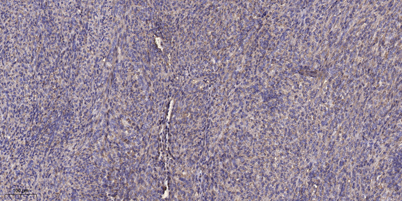
| Dilution | WB 1:500-2000;IHC-p 1:50-300; ELISA 2000-20000 |
| Purification | The antibody was affinity-purified from rabbit antiserum by affinity-chromatography using epitope-specific immunogen. |
| Concentration | 1 mg/ml |
| Storage stability | -20°C/1 year |
| Molecular Weight (Da) | |
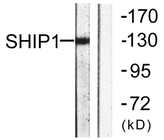
| Observed band (KD) | 133kD |
| Background | This gene is a member of the inositol polyphosphate-5-phosphatase (INPP5) family and encodes a protein with an N-terminal SH2 domain, an inositol phosphatase domain, and two C-terminal protein interaction domains. Expression of this protein is restricted to hematopoietic cells where its movement from the cytosol to the plasma membrane is mediated by tyrosine phosphorylation. At the plasma membrane, the protein hydrolyzes the 5' phosphate from phosphatidylinositol (3,4,5)-trisphosphate and inositol-1,3,4,5-tetrakisphosphate, thereby affecting multiple signaling pathways. The protein is also partly localized to the nucleus, where it may be involved in nuclear inositol phosphate signaling processes. Overall, the protein functions as a negative regulator of myeloid cell proliferation and survival. Mutations in this gene are associated with defects and cancers of the immune system. A |
| Function | catalytic activity:Phosphatidylinositol 3,4,5-trisphosphate + H(2)O = phosphatidylinositol 3,4-bisphosphate + phosphate.,domain:The NPXY sequence motif found in many tyrosine-phosphorylated proteins is required for the specific binding of the PID domain.,domain:The SH2 domain interacts with tyrosine phosphorylated forms of proteins such as SHC1 or PTPN11/SHP-2. It competes with that of GRB2 for binding to phosphorylated SHC1 to inhibit the Ras pathway. It is also required for tyrosine phosphorylation.,enzyme regulation:Activated upon translocation to the sites of synthesis of PtdIns(3,4,5)P3 in the membrane.,function:Phosphatidylinositol (PtdIns) phosphatase that specifically hydrolyzes the 5-phosphate of phosphatidylinositol-3,4,5-trisphosphate (PtdIns(3,4,5)P3) to produce PtdIns(3,4)P2, thereby negatively regulating the PI3K (phosphoinositide 3-kinase) pathways. Acts as a negative regu |
| Subcellular location | Cytoplasm . Cell membrane ; Peripheral membrane protein . Membrane raft . Cytoplasm, cytoskeleton . Membrane ; Peripheral membrane protein . Translocates to the plasma membrane when activated, translocation is probably due to different mechanisms depending on the stimulus and cell type. Translocates from the cytoplasm to membrane ruffles in a FCGR3/CD16-dependent manner. Colocalizes with FC-gamma-RIIB receptor (FCGR2B) or FCGR3/CD16 at membrane ruffles. Tyrosine phosphorylation may also participate in membrane localization. . |
| Expression | Specifically expressed in immune and hematopoietic cells. Expressed in bone marrow and blood cells. Levels vary considerably within this compartment. Present in at least 74% of immature CD34+ cells, whereas within the more mature population of CD33+ cells, it is present in only 10% of cells. Present in the majority of T-cells, while it is present in a minority of B-cells (at protein level). |

Use collapsible tabs for more detailed information that will help customers make a purchasing decision.
Ex: Shipping and return policies, size guides, and other common questions.