 Français
 English
IRAK-1 (phospho Thr209) rabbit pAb
AO-06-ES5914-50

| ELK.No | ES5914 |
| Product name | IRAK-1 (phospho Thr209) rabbit pAb |
| Reactivity | Human;Rat;Mouse; |
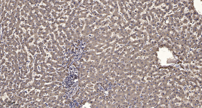
| Applications | WB;ELISA;IHC |
| Other name | IRAK1; IRAK; Interleukin-1 receptor-associated kinase 1; IRAK-1 |
| Size | 50μL |
| Unit price ($) | 148 |
| Human gene ID | 3654 |
| Human Swiss-Prot | P51617 |
| Source | Rabbit |
| Isotype | IgG |
| Target | IRAK-1 |
| Fields | >>MAPK signaling pathway;>>NF-kappa B signaling pathway;>>Toll-like receptor signaling pathway;>>Neurotrophin signaling pathway;>>Alcoholic liver disease;>>Pathogenic Escherichia coli infection;>>Salmonella infection;>>Pertussis;>>Yersinia infection;>>Leishmaniasis;>>Chagas disease;>>Toxoplasmosis;>>Tuberculosis;>>Hepatitis B;>>Measles;>>Herpes simplex virus 1 infection;>>Epstein-Barr virus infection;>>Human immunodeficiency virus 1 infection;>>Coronavirus disease - COVID-19;>>Lipid and atherosclerosis |
| Gene name | IRAK1 |
| Protein name | Interleukin-1 receptor-associated kinase 1 |
| Human gene link | View Human Gene |
| Human Swiss link | View Human Swiss-Prot |
| Mouse gene ID | |
| Mouse gene link | |
| Mouse Swiss-Prot | Q62406 |
| Mouse Swiss link | View Mouse Swiss-Prot |
| Rat gene ID | |
| Rat gene link | |
| Rat Swiss-Prot | |
| Rat Swiss link | |
| Immunogen | The antiserum was produced against synthesized peptide derived from human IRAK1 around the phosphorylation site of Thr209. AA range:175-224 |
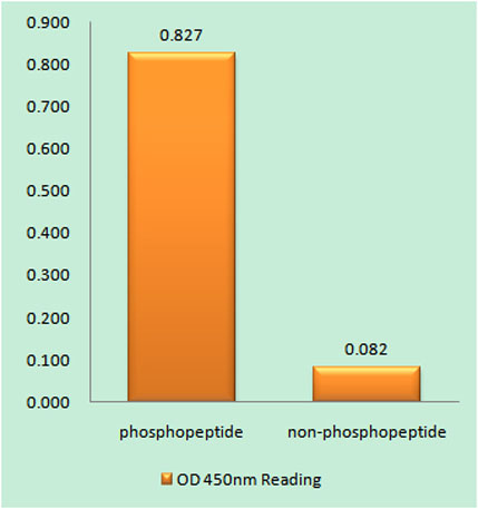
| Specificity | Phospho-IRAK-1 (T209) Polyclonal Antibody detects endogenous levels of IRAK-1 protein only when phosphorylated at T209. |
| Formulation | Liquid in PBS containing 50% glycerol, 0.5% BSA and 0.02% sodium azide. |
| Clonality | Polyclonal |
| Dilution | WB 1:500-2000;IHC-p 1:50-300; ELISA 2000-20000 |
| Purification | The antibody was affinity-purified from rabbit antiserum by affinity-chromatography using epitope-specific immunogen. |
| Concentration | 1 mg/ml |
| Storage stability | -20°C/1 year |
| Molecular Weight (Da) | |
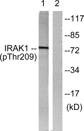
| Observed band (KD) | 77kD |
| Background | This gene encodes the interleukin-1 receptor-associated kinase 1, one of two putative serine/threonine kinases that become associated with the interleukin-1 receptor (IL1R) upon stimulation. This gene is partially responsible for IL1-induced upregulation of the transcription factor NF-kappa B. Alternatively spliced transcript variants encoding different isoforms have been found for this gene. [provided by RefSeq, Jul 2008], |
| Function | catalytic activity:ATP + a protein = ADP + a phosphoprotein.,cofactor:Magnesium.,function:Binds to the IL-1 type I receptor following IL-1 engagement, triggering intracellular signaling cascades leading to transcriptional up-regulation and mRNA stabilization. Isoform 1 binds rapidly but is then degraded allowing isoform 2 to mediate a slower, more sustained response to the cytokine. Isoform 2 is inactive suggesting that the kinase activity of this enzyme is not required for IL-1 signaling. Once phosphorylated, IRAK1 recruits the adapter protein PELI1.,PTM:Autophosphorylated or is transphosphorylated by IRAK4 following recruitment to the IL-1RI. In the case of isoform 1, this is linked to ubiquitination and degradation.,similarity:Belongs to the protein kinase superfamily.,similarity:Belongs to the protein kinase superfamily. TKL Ser/Thr protein kinase family. Pelle subfamily.,similarity: |
| Subcellular location | Cytoplasm . Nucleus . Lipid droplet . Translocates to the nucleus when sumoylated. RSAD2/viperin recruits it to the lipid droplet (By similarity). . |
| Expression | Isoform 1 and isoform 2 are ubiquitously expressed in all tissues examined, with isoform 1 being more strongly expressed than isoform 2. |

Use collapsible tabs for more detailed information that will help customers make a purchasing decision.
Ex: Shipping and return policies, size guides, and other common questions.