 Français
 English
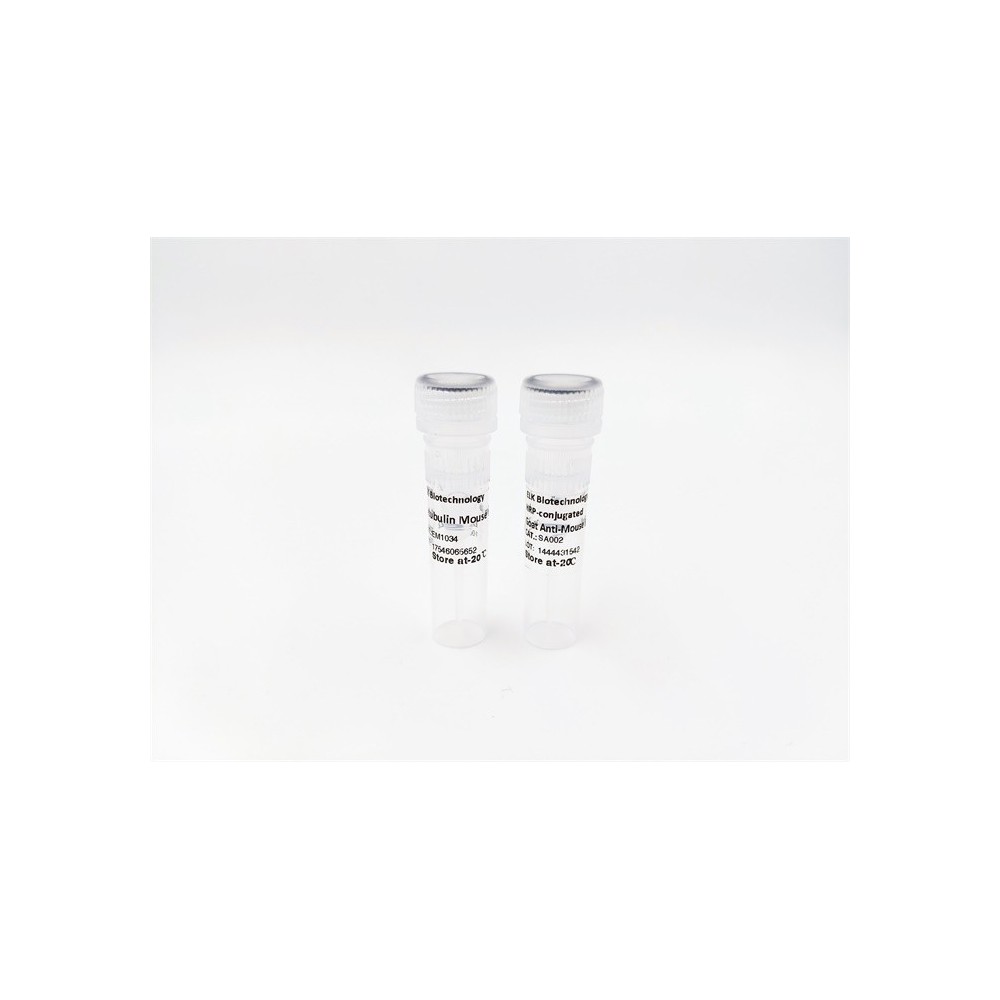
IRF-3 (phospho Ser386) rabbit pAb
AO-06-ES5922-100

| ELK.No | ES5922 |
| Product name | IRF-3 (phospho Ser386) rabbit pAb |
| Reactivity | Human;Rat;Mouse; |
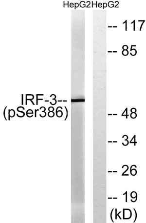
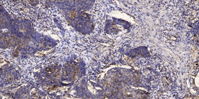
| Applications | WB;IHC |
| Other name | IRF3; Interferon regulatory factor 3; IRF-3 |
| Size | 100μL |
| Unit price ($) | 248 |
| Human gene ID | 3661 |
| Human Swiss-Prot | Q14653 |
| Source | Rabbit |
| Isotype | IgG |
| Target | IRF-3 |
| Fields | >>Toll-like receptor signaling pathway;>>NOD-like receptor signaling pathway;>>RIG-I-like receptor signaling pathway;>>Cytosolic DNA-sensing pathway;>>Alcoholic liver disease;>>Shigellosis;>>Pertussis;>>Yersinia infection;>>Hepatitis C;>>Hepatitis B;>>Measles;>>Human cytomegalovirus infection;>>Influenza A;>>Human papillomavirus infection;>>Kaposi sarcoma-associated herpesvirus infection;>>Herpes simplex virus 1 infection;>>Epstein-Barr virus infection;>>Human immunodeficiency virus 1 infection;>>Coronavirus disease - COVID-19;>>Viral carcinogenesis;>>Lipid and atherosclerosis |
| Gene name | IRF3 |
| Protein name | Interferon regulatory factor 3 |
| Human gene link | View Human Gene |
| Human Swiss link | View Human Swiss-Prot |
| Mouse gene ID | |
| Mouse gene link | |
| Mouse Swiss-Prot | P70671 |
| Mouse Swiss link | View Mouse Swiss-Prot |
| Rat gene ID | |
| Rat gene link | |
| Rat Swiss-Prot | |
| Rat Swiss link | |
| Immunogen | The antiserum was produced against synthesized peptide derived from human IRF-3 around the phosphorylation site of Ser386. AA range:352-401 |
| Specificity | Phospho-IRF-3 (S386) Polyclonal Antibody detects endogenous levels of IRF-3 protein only when phosphorylated at S386. |
| Formulation | Liquid in PBS containing 50% glycerol, 0.5% BSA and 0.02% sodium azide. |
| Clonality | Polyclonal |
| Dilution | WB 1:500-2000;IHC-p 1:50-300 |
| Purification | The antibody was affinity-purified from rabbit antiserum by affinity-chromatography using epitope-specific immunogen. |
| Concentration | 1 mg/ml |
| Storage stability | -20°C/1 year |
| Molecular Weight (Da) | |
| Observed band (KD) | 48-55kd |
| Background | This gene encodes a member of the interferon regulatory transcription factor (IRF) family. The encoded protein is found in an inactive cytoplasmic form that upon serine/threonine phosphorylation forms a complex with CREBBP. This complex translocates to the nucleus and activates the transcription of interferons alpha and beta, as well as other interferon-induced genes. Alternatively spliced transcript variants encoding multiple isoforms have been observed for this gene. [provided by RefSeq, Nov 2011], |
| Function | function:Mediates interferon-stimulated response element (ISRE) promoter activation. Functions as a molecular switch for antiviral activity. DsRNA generated during the course of an viral infection leads to IRF3 phosphorylation on the C-terminal serine/threonine cluster. This induces a conformational change, leading to its dimerization, nuclear localization and association with CREB binding protein (CREBBP) to form dsRNA-activated factor 1 (DRAF1), a complex which activates the transcription of genes under the control of ISRE. The complex binds to the IE and PRDIII regions on the IFN-alpha and IFN-beta promoters respectively. IRF-3 does not have any transcription activation domains.,PTM:Constitutively phosphorylated on many serines residues. C-terminal serine/threonine cluster is phosphorylated in response of induction by IKBKE and TBK1. Ser-385 and Ser-386 may be specifically phosphoryla |
| Subcellular location | Cytoplasm . Nucleus . Mitochondrion . Shuttles between cytoplasmic and nuclear compartments, with export being the prevailing effect (PubMed:10805757). When activated, IRF3 interaction with CREBBP prevents its export to the cytoplasm (PubMed:10805757). Recruited to mitochondria via TOMM70:HSP90AA1 upon Sendai virus infection (PubMed:25609812). . |
| Expression | Expressed constitutively in a variety of tissues. |

Use collapsible tabs for more detailed information that will help customers make a purchasing decision.
Ex: Shipping and return policies, size guides, and other common questions.