 Français
 English
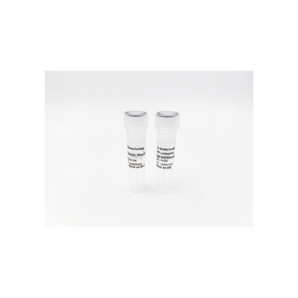

| ELK.No | ES6033 |
| Product name | Cytokeratin 14/16 rabbit pAb |
| Reactivity | Human;Mouse;Rat |
| Applications | WB;IHC;IF;ELISA |
| Other name | KRT14; Keratin; type I cytoskeletal 14; Cytokeratin-14; CK-14; Keratin-14; K14; KRT16; KRT16A; Keratin, type I cytoskeletal 16; Cytokeratin-16; CK-16; Keratin-16; K16 |
| Size | 100μL |
| Unit price ($) | 248 |
| Human gene ID | 3861/3868/3872 |
| Human Swiss-Prot | P02533/P08779 |
| Source | Rabbit |
| Isotype | IgG |
| Target | Cytokeratin 14/16 |
| Fields | >>Estrogen signaling pathway;>>Staphylococcus aureus infection |
| Gene name | KRT14/KRT16 |
| Protein name | Keratin type I cytoskeletal 14/16 |
| Human gene link | View Human Gene |
| Human Swiss link | View Human Swiss-Prot |
| Mouse gene ID | 16664/16666 |
| Mouse gene link | View Mouse Gene |
| Mouse Swiss-Prot | |
| Mouse Swiss link | |
| Rat gene ID | 287701 |
| Rat gene link | View Rat Gene |
| Rat Swiss-Prot | Q6IFV1 |
| Rat Swiss link | View Rat Swiss-Prot |
| Immunogen | The antiserum was produced against synthesized peptide derived from human Keratin 14. AA range:1-50 |
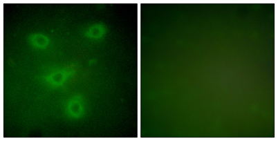
| Specificity | Cytokeratin 14/16 Polyclonal Antibody detects endogenous levels of Cytokeratin 14/16 protein. |
| Formulation | Liquid in PBS containing 50% glycerol, 0.5% BSA and 0.02% sodium azide. |
| Clonality | Polyclonal |
| Dilution | Western Blot: 1/500 - 1/2000. Immunohistochemistry: 1/100 - 1/300. Immunofluorescence: 1/200 - 1/1000. ELISA: 1/20000. Not yet tested in other applications. |
| Purification | The antibody was affinity-purified from rabbit antiserum by affinity-chromatography using epitope-specific immunogen. |
| Concentration | 1 mg/ml |
| Storage stability | -20°C/1 year |
| Molecular Weight (Da) | |
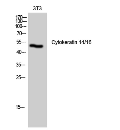
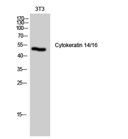
| Observed band (KD) | 52kD |
| Background | This gene encodes a member of the keratin family, the most diverse group of intermediate filaments. This gene product, a type I keratin, is usually found as a heterotetramer with two keratin 5 molecules, a type II keratin. Together they form the cytoskeleton of epithelial cells. Mutations in the genes for these keratins are associated with epidermolysis bullosa simplex. At least one pseudogene has been identified at 17p12-p11. [provided by RefSeq, Jul 2008], |
| Function | disease:Defects in KRT14 are a cause of epidermolysis bullosa simplex Dowling-Meara type (DM-EBS) [MIM:131760]. DM-EBS is a severe form of intraepidermal epidermolysis bullosa characterized by generalized herpetiform blistering, milia formation, dystrophic nails, and mucous membrane involvement.,disease:Defects in KRT14 are a cause of epidermolysis bullosa simplex Koebner type (K-EBS) [MIM:131900]. K-EBS is a form of intraepidermal epidermolysis bullosa characterized by generalized skin blistering. The phenotype is not fundamentally distinct from the Dowling-Meara type, althought it is less severe.,disease:Defects in KRT14 are a cause of epidermolysis bullosa simplex Weber-Cockayne type (WC-EBS) [MIM:131800]. WC-EBS is a form of intraepidermal epidermolysis bullosa characterized by blistering limited to palmar and plantar areas of the skin.,disease:Defects in KRT14 are the cause of derma |
| Subcellular location | Cytoplasm. Nucleus. Expressed in both as a filamentous pattern. |
| Expression | Expressed in the corneal epithelium (at protein level) (PubMed:26758872). Detected in the basal layer, lowered within the more apically located layers specifically in the stratum spinosum, stratum granulosum but is not detected in stratum corneum. Strongly expressed in the outer root sheath of anagen follicles but not in the germinative matrix, inner root sheath or hair (PubMed:9457912). Found in keratinocytes surrounding the club hair during telogen (PubMed:9457912). |

Use collapsible tabs for more detailed information that will help customers make a purchasing decision.
Ex: Shipping and return policies, size guides, and other common questions.