 Français
 English
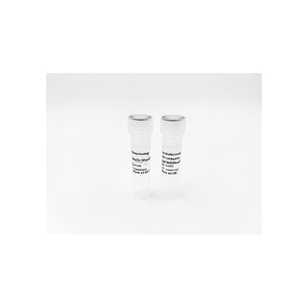

| ELK.No | ES6077 |
| Product name | Laminin α-5 rabbit pAb |
| Reactivity | Human;Mouse |
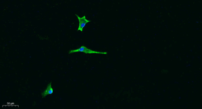
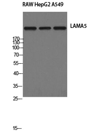
| Applications | WB;IHC;IF;ELISA |
| Other name | LAMA5; KIAA0533; KIAA1907; Laminin subunit alpha-5; Laminin-10 subunit alpha; Laminin-11 subunit alpha; Laminin-15 subunit alpha |
| Size | 50μL |
| Unit price ($) | 148 |
| Human gene ID | 3911 |
| Human Swiss-Prot | O15230 |
| Source | Rabbit |
| Isotype | IgG |
| Target | LAMA5 |
| Fields | >>PI3K-Akt signaling pathway;>>Focal adhesion;>>ECM-receptor interaction;>>Toxoplasmosis;>>Amoebiasis;>>Human papillomavirus infection;>>Pathways in cancer;>>Small cell lung cancer |
| Gene name | LAMA5 |
| Protein name | Laminin subunit alpha-5 |
| Human gene link | View Human Gene |
| Human Swiss link | View Human Swiss-Prot |
| Mouse gene ID | 16776 |
| Mouse gene link | View Mouse Gene |
| Mouse Swiss-Prot | Q61001 |
| Mouse Swiss link | View Mouse Swiss-Prot |
| Rat gene ID | |
| Rat gene link | |
| Rat Swiss-Prot | |
| Rat Swiss link | |
| Immunogen | The antiserum was produced against synthesized peptide derived from human LAMA5. AA range:2381-2430 |
| Specificity | Laminin α-5 Polyclonal Antibody detects endogenous levels of Laminin α-5 protein. |
| Formulation | Liquid in PBS containing 50% glycerol, 0.5% BSA and 0.02% sodium azide. |
| Clonality | Polyclonal |
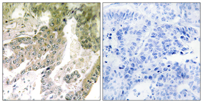
| Dilution | Western Blot: 1/500 - 1/2000. IHC-p: 1:100-300 ELISA: 1/20000. IF 1:100-300 Not yet tested in other applications. |
| Purification | The antibody was affinity-purified from rabbit antiserum by affinity-chromatography using epitope-specific immunogen. |
| Concentration | 1 mg/ml |
| Storage stability | -20°C/1 year |
| Molecular Weight (Da) | |
| Observed band (KD) | 400kD |
| Background | This gene encodes one of the vertebrate laminin alpha chains. Laminins, a family of extracellular matrix glycoproteins, are the major noncollagenous constituent of basement membranes. They have been implicated in a wide variety of biological processes including cell adhesion, differentiation, migration, signaling, neurite outgrowth and metastasis. Laminins are composed of 3 non identical chains: laminin alpha, beta and gamma (formerly A, B1, and B2, respectively) and they form a cruciform structure consisting of 3 short arms, each formed by a different chain, and a long arm composed of all 3 chains. Each laminin chain is a multidomain protein encoded by a distinct gene. The protein encoded by this gene is the alpha-5 subunit of of laminin-10 (laminin-511), laminin-11 (laminin-521) and laminin-15 (laminin-523). [provided by RefSeq, Jun 2013], |
| Function | domain:Domain G is globular and is part of the major cell-binding site located in the long arm of the laminin heterotrimer.,function:Binding to cells via a high affinity receptor, laminin is thought to mediate the attachment, migration and organization of cells into tissues during embryonic development by interacting with other extracellular matrix components.,similarity:Contains 1 laminin IV type A domain.,similarity:Contains 1 laminin N-terminal domain.,similarity:Contains 22 laminin EGF-like domains.,similarity:Contains 5 laminin G-like domains.,subcellular location:Major component.,subunit:Laminin-15 complex is an heterotrimer composed of three chains (alpha-5/beta-2/gamma-3) which are bound to each other by disulfide bonds into a cross-shaped molecule comprising one long and three short arms with globules at each end.,tissue specificity:Expressed in heart, lung, kidney, skeletal mus |
| Subcellular location | Secreted, extracellular space, extracellular matrix, basement membrane. Major component. |
| Expression | Expressed in heart, lung, kidney, skeletal muscle, pancreas, retina and placenta. Little or no expression in brain and liver. |

Use collapsible tabs for more detailed information that will help customers make a purchasing decision.
Ex: Shipping and return policies, size guides, and other common questions.