 Français
 English

| ELK.No | ES6247 |
| Product name | MLF1 rabbit pAb |
| Reactivity | Human;Mouse |
| Applications | WB;IHC |
| Other name | MLF1; Myeloid leukemia factor 1; Myelodysplasia-myeloid leukemia factor 1 |
| Size | 50μL |
| Unit price ($) | 148 |
| Human gene ID | 4291 |
| Human Swiss-Prot | P58340 |
| Source | Rabbit |
| Isotype | IgG |
| Target | MLF1 |
| Fields | >>Transcriptional misregulation in cancer |
| Gene name | MLF1 |
| Protein name | Myeloid leukemia factor 1 |
| Human gene link | View Human Gene |
| Human Swiss link | View Human Swiss-Prot |
| Mouse gene ID | 17349 |
| Mouse gene link | View Mouse Gene |
| Mouse Swiss-Prot | Q9QWV4 |
| Mouse Swiss link | View Mouse Swiss-Prot |
| Rat gene ID | |
| Rat gene link | |
| Rat Swiss-Prot | |
| Rat Swiss link | |
| Immunogen | Synthesized peptide derived from MLF1 . at AA range: 130-210 |
| Specificity | MLF1 Polyclonal Antibody detects endogenous levels of MLF1 protein. |
| Formulation | Liquid in PBS containing 50% glycerol, 0.5% BSA and 0.02% sodium azide. |
| Clonality | Polyclonal |
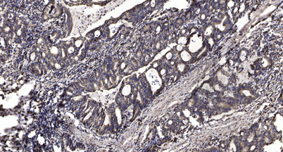
| Dilution | WB 1:500-2000;IHC-p 1:50-300 |
| Purification | The antibody was affinity-purified from rabbit antiserum by affinity-chromatography using epitope-specific immunogen. |
| Concentration | 1 mg/ml |
| Storage stability | -20°C/1 year |
| Molecular Weight (Da) | |
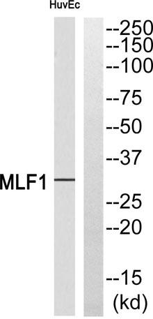
| Observed band (KD) | 30kD |
| Background | myeloid leukemia factor 1(MLF1) Homo sapiens This gene encodes an oncoprotein which is thought to play a role in the phenotypic determination of hemopoetic cells. Translocations between this gene and nucleophosmin have been associated with myelodysplastic syndrome and acute myeloid leukemia. Multiple transcript variants encoding different isoforms have been found for this gene. [provided by RefSeq, Sep 2010], |
| Function | disease:A chromosomal aberration involving MLF1 is a cause of myelodysplastic syndrome (MDS). Translocation t(3;5)(q25.1;q34) with NPM1/NPM.,function:Involved in lineage commitment of primary hemopoietic progenitors by restricting erythroid formation and enhancing myeloid formation. Interferes with erythopoietin-induced erythroid terminal differentiation by preventing cells from exiting the cell cycle through suppression of CDKN1B/p27Kip1 levels. Suppresses RFWD2/COP1 activity via CSN3 which activates p53 and induces cell cycle arrest. Binds DNA and affects the expression of a number of genes so may function as a transcription factor in the nucleus.,PTM:Phosphorylation is required for binding to YWHAZ.,similarity:Belongs to the MLF family.,subcellular location:In non-hematopoietic cells, resides primarily in the cytoplasm with some punctate nuclear localization. Shuttles between the cyto |
| Subcellular location | Cytoplasm . Nucleus . Cell projection, cilium . Cytoplasm, cytoskeleton, cilium basal body . Shuttles between the cytoplasm and nucleus. . |
| Expression | Most abundant in testis, ovary, skeletal muscle, heart, kidney and colon. Low expression in spleen, thymus and peripheral blood leukocytes. |

Use collapsible tabs for more detailed information that will help customers make a purchasing decision.
Ex: Shipping and return policies, size guides, and other common questions.