 Français
 English

| ELK.No | ES6301 |
| Product name | MYBPC1 rabbit pAb |
| Reactivity | Human;Mouse;Rat |
| Applications | WB;ELISA;IHC |
| Other name | MYBPC1; MYBPCS; Myosin-binding protein C; slow-type; Slow MyBP-C; C-protein, skeletal muscle slow isoform |
| Size | 50μL |
| Unit price ($) | 148 |
| Human gene ID | 4604 |
| Human Swiss-Prot | Q00872 |
| Source | Rabbit |
| Isotype | IgG |
| Target | MYBPC1 |
| Fields | |
| Gene name | MYBPC1 |
| Protein name | Myosin-binding protein C slow-type |
| Human gene link | View Human Gene |
| Human Swiss link | View Human Swiss-Prot |
| Mouse gene ID | |
| Mouse gene link | |
| Mouse Swiss-Prot | |
| Mouse Swiss link | |
| Rat gene ID | 362867 |
| Rat gene link | View Rat Gene |
| Rat Swiss-Prot | Q63518 |
| Rat Swiss link | View Rat Swiss-Prot |
| Immunogen | The antiserum was produced against synthesized peptide derived from human MYBPC1. AA range:218-267 |
| Specificity | MYBPC1 Polyclonal Antibody detects endogenous levels of MYBPC1 protein. |
| Formulation | Liquid in PBS containing 50% glycerol, 0.5% BSA and 0.02% sodium azide. |
| Clonality | Polyclonal |
| Dilution | WB 1:500-2000;IHC-p 1:50-300; ELISA 2000-20000 |
| Purification | The antibody was affinity-purified from rabbit antiserum by affinity-chromatography using epitope-specific immunogen. |
| Concentration | 1 mg/ml |
| Storage stability | -20°C/1 year |
| Molecular Weight (Da) | |
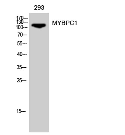
| Observed band (KD) | 120kD |
| Background | This gene encodes a member of the myosin-binding protein C family. Myosin-binding protein C family members are myosin-associated proteins found in the cross-bridge-bearing zone (C region) of A bands in striated muscle. The encoded protein is the slow skeletal muscle isoform of myosin-binding protein C and plays an important role in muscle contraction by recruiting muscle-type creatine kinase to myosin filaments. Mutations in this gene are associated with distal arthrogryposis type I. Alternatively spliced transcript variants encoding multiple isoforms have been observed for this gene. [provided by RefSeq, Dec 2011], |
| Function | function:Thick filament-associated protein located in the crossbridge region of vertebrate striated muscle a bands. In vitro it binds MHC, F-actin and native thin filaments, and modifies the activity of actin-activated myosin ATPase. It may modulate muscle contraction or may play a more structural role.,similarity:Belongs to the immunoglobulin superfamily. MyBP family.,similarity:Contains 3 fibronectin type-III domains.,similarity:Contains 7 Ig-like C2-type (immunoglobulin-like) domains., |
| Subcellular location | cytosol,myofibril,myosin filament, |
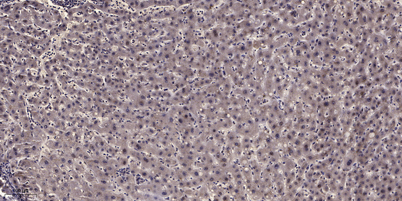
| Expression | Brain,Fetal skeletal muscle,Hippocampus,Liver,Muscle pool- 2 tissue |

Use collapsible tabs for more detailed information that will help customers make a purchasing decision.
Ex: Shipping and return policies, size guides, and other common questions.