 Français
 English

| ELK.No | ES6545 |
| Product name | PDGFR-β rabbit pAb |
| Reactivity | Human;Mouse;Rat |
| Applications | WB;IHC;IF;ELISA |
| Other name | PDGFRB; PDGFR; PDGFR1; Platelet-derived growth factor receptor beta; PDGF-R-beta; PDGFR-beta; Beta platelet-derived growth factor receptor; Beta-type platelet-derived growth factor receptor; CD140 antigen-like family member B; Platelet-deri |
| Size | 100μL |
| Unit price ($) | 248 |
| Human gene ID | 5159 |
| Human Swiss-Prot | P09619 |
| Source | Rabbit |
| Isotype | IgG |
| Target | PDGFR-β |
| Fields | >>EGFR tyrosine kinase inhibitor resistance;>>MAPK signaling pathway;>>Ras signaling pathway;>>Rap1 signaling pathway;>>Calcium signaling pathway;>>Phospholipase D signaling pathway;>>PI3K-Akt signaling pathway;>>Focal adhesion;>>Gap junction;>>JAK-STAT signaling pathway;>>Regulation of actin cytoskeleton;>>Human papillomavirus infection;>>Pathways in cancer;>>MicroRNAs in cancer;>>Glioma;>>Prostate cancer;>>Melanoma;>>Central carbon metabolism in cancer;>>Choline metabolism in cancer |
| Gene name | PDGFRB |
| Protein name | Platelet-derived growth factor receptor beta |
| Human gene link | View Human Gene |
| Human Swiss link | View Human Swiss-Prot |
| Mouse gene ID | 18596 |
| Mouse gene link | View Mouse Gene |
| Mouse Swiss-Prot | P05622 |
| Mouse Swiss link | View Mouse Swiss-Prot |
| Rat gene ID | 24629 |
| Rat gene link | View Rat Gene |
| Rat Swiss-Prot | Q05030 |
| Rat Swiss link | View Rat Swiss-Prot |
| Immunogen | The antiserum was produced against synthesized peptide derived from human PDGFR beta. AA range:991-1040 |
| Specificity | PDGFR-β Polyclonal Antibody detects endogenous levels of PDGFR-β protein. |
| Formulation | Liquid in PBS containing 50% glycerol, 0.5% BSA and 0.02% sodium azide. |
| Clonality | Polyclonal |
| Dilution | Western Blot: 1/500 - 1/2000. Immunohistochemistry: 1/100 - 1/300. Immunofluorescence: 1/200 - 1/1000. ELISA: 1/5000. Not yet tested in other applications. |
| Purification | The antibody was affinity-purified from rabbit antiserum by affinity-chromatography using epitope-specific immunogen. |
| Concentration | 1 mg/ml |
| Storage stability | -20°C/1 year |
| Molecular Weight (Da) | |
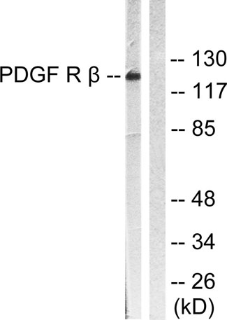
| Observed band (KD) | 135-180kD |
| Background | This gene encodes a cell surface tyrosine kinase receptor for members of the platelet-derived growth factor family. These growth factors are mitogens for cells of mesenchymal origin. The identity of the growth factor bound to a receptor monomer determines whether the functional receptor is a homodimer or a heterodimer, composed of both platelet-derived growth factor receptor alpha and beta polypeptides. This gene is flanked on chromosome 5 by the genes for granulocyte-macrophage colony-stimulating factor and macrophage-colony stimulating factor receptor; all three genes may be implicated in the 5-q syndrome. A translocation between chromosomes 5 and 12, that fuses this gene to that of the translocation, ETV6, leukemia gene, results in chronic myeloproliferative disorder with eosinophilia. [provided by RefSeq, Jul 2008], |
| Function | catalytic activity:ATP + a [protein]-L-tyrosine = ADP + a [protein]-L-tyrosine phosphate.,disease:A chromosomal aberration involving PDGFRB is a cause in many instances of chronic myeloproliferative disorder with eosinophilia (MPE) [MIM:131440]. Translocation t(5;12) with ETV6 on chromosome 12 creating an PDGFRB-ETV6 fusion protein.,disease:A chromosomal aberration involving PDGFRB is found in a form of chronic myelomonocytic leukemia (CMML). Translocation t(5;12)(q33;p13) with EVT6/TEL. It is characterized by abnormal clonal myeloid proliferation and by progression to acute myelogenous leukemia (AML).,disease:A chromosomal aberration involving PDGFRB may be a cause of acute myelogenous leukemia. Translocation t(5;14)(q33;q32) with TRIP11. The fusion protein may be involved in clonal evolution of leukemia and eosinophilia.,disease:A chromosomal aberration involving PDGFRB may be a cause |
| Subcellular location | Cell membrane; Single-pass type I membrane protein. Cytoplasmic vesicle. Lysosome lumen. After ligand binding, the autophosphorylated receptor is ubiquitinated and internalized, leading to its degradation. |
| Expression | Brain,Spleen, |

Use collapsible tabs for more detailed information that will help customers make a purchasing decision.
Ex: Shipping and return policies, size guides, and other common questions.