 Français
 English
CD31 (phospho Tyr713) rabbit pAb
AO-06-ES6563-100

| ELK.No | ES6563 |
| Product name | CD31 (phospho Tyr713) rabbit pAb |
| Reactivity | Human;Mouse |
| Applications | WB;IHC;IF;ELISA |
| Other name | PECAM1; Platelet endothelial cell adhesion molecule; PECAM-1; EndoCAM; GPIIA'; PECA1; CD antigen CD31 |
| Size | 100μL |
| Unit price ($) | 248 |
| Human gene ID | 5175 |
| Human Swiss-Prot | P16284 |
| Source | Rabbit |
| Isotype | IgG |
| Target | CD31 |
| Fields | >>Cell adhesion molecules;>>Leukocyte transendothelial migration;>>Malaria;>>Fluid shear stress and atherosclerosis |
| Gene name | PECAM1 |
| Protein name | Platelet endothelial cell adhesion molecule |
| Human gene link | View Human Gene |
| Human Swiss link | View Human Swiss-Prot |
| Mouse gene ID | 18613 |
| Mouse gene link | View Mouse Gene |
| Mouse Swiss-Prot | Q08481 |
| Mouse Swiss link | View Mouse Swiss-Prot |
| Rat gene ID | |
| Rat gene link | |
| Rat Swiss-Prot | |
| Rat Swiss link | |
| Immunogen | The antiserum was produced against synthesized peptide derived from human PECAM-1 around the phosphorylation site of Tyr713. AA range:686-735 |
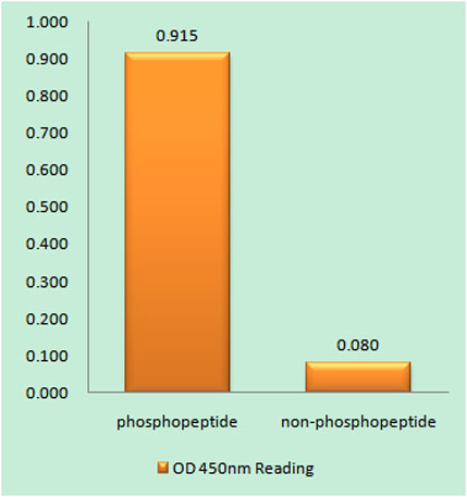
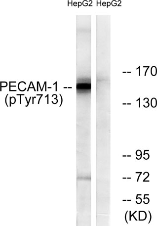
| Specificity | Phospho-CD31 (Y713) Polyclonal Antibody detects endogenous levels of CD31 protein only when phosphorylated at Y713. |
| Formulation | Liquid in PBS containing 50% glycerol, 0.5% BSA and 0.02% sodium azide. |
| Clonality | Polyclonal |
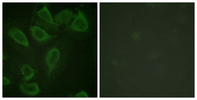
| Dilution | Western Blot: 1/500 - 1/2000. Immunohistochemistry: 1/100 - 1/300. Immunofluorescence: 1/200 - 1/1000. ELISA: 1/20000. Not yet tested in other applications. |
| Purification | The antibody was affinity-purified from rabbit antiserum by affinity-chromatography using epitope-specific immunogen. |
| Concentration | 1 mg/ml |
| Storage stability | -20°C/1 year |
| Molecular Weight (Da) | |
| Observed band (KD) | 150kD |
| Background | The protein encoded by this gene is found on the surface of platelets, monocytes, neutrophils, and some types of T-cells, and makes up a large portion of endothelial cell intercellular junctions. The encoded protein is a member of the immunoglobulin superfamily and is likely involved in leukocyte migration, angiogenesis, and integrin activation. [provided by RefSeq, May 2010], |
| Function | function:This protein is a cell adhesion molecule expressed on platelets and at endothelial cell intercellular junctions.,online information:CD31 entry,online information:PECAM-1,online information:The Singapore human mutation and polymorphism database,PTM:Phosphorylated on Ser and Tyr residues after cellular activation.,similarity:Contains 6 Ig-like C2-type (immunoglobulin-like) domains.,tissue specificity:Long isoform predominates all tissues examined, isoform Delta12 was detected only in trachea and isoform Delta14-15 only in lung, isoform Delta14 was detected in all tissues examined with the strongest expression in heart., |
| Subcellular location | Cell membrane ; Single-pass type I membrane protein . Cell surface expression on neutrophils is down-regulated upon fMLP or CXCL8/IL8-mediated stimulation. .; [Isoform Long]: Cell membrane ; Single-pass type I membrane protein . Membrane raft . Cell junction . Localizes to the lateral border recycling compartment (LBRC) and recycles from the LBRC to the junction in resting endothelial cells. .; [Isoform Delta15]: Cell junction . Localizes to the lateral border recycling compartment (LBRC) and recycles from the LBRC to the junction in resting endothelial cells. |
| Expression | Expressed on platelets and leukocytes and is primarily concentrated at the borders between endothelial cells (PubMed:18388311, PubMed:21464369). Expressed in human umbilical vein endothelial cells (HUVECs) (at protein level) (PubMed:19342684, PubMed:17580308). Expressed on neutrophils (at protein level) (PubMed:17580308). Isoform Long predominates in all tissues examined (PubMed:12433657). Isoform Delta12 is detected only in trachea (PubMed:12433657). Isoform Delta14-15 is only detected in lung (PubMed:12433657). Isoform Delta14 is detected in all tissues examined with the strongest expression in heart (PubMed:12433657). Isoform Delta15 is expressed in brain, testis, ovary, cell surface of platelets, human umbilical vein endothelial cells (HUVECs), Jurkat T-cell leukemia, human erythroleuk |

Use collapsible tabs for more detailed information that will help customers make a purchasing decision.
Ex: Shipping and return policies, size guides, and other common questions.