 Français
 English

| ELK.No | ES6567 |
| Product name | PEG3 rabbit pAb |
| Reactivity | Human;Rat;Mouse; |
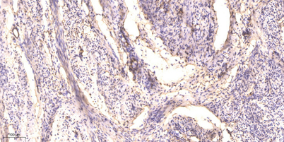
| Applications | WB;IHC |
| Other name | PEG3; KIAA0287; ZSCAN24; Paternally-expressed gene 3 protein; Zinc finger and SCAN domain-containing protein 24 |
| Size | 50μL |
| Unit price ($) | 148 |
| Human gene ID | 5178 |
| Human Swiss-Prot | Q9GZU2 |
| Source | Rabbit |
| Isotype | IgG |
| Target | PEG3 |
| Fields | |
| Gene name | PEG3 |
| Protein name | Paternally-expressed gene 3 protein |
| Human gene link | View Human Gene |
| Human Swiss link | View Human Swiss-Prot |
| Mouse gene ID | |
| Mouse gene link | |
| Mouse Swiss-Prot | Q3URU2 |
| Mouse Swiss link | View Mouse Swiss-Prot |
| Rat gene ID | |
| Rat gene link | |
| Rat Swiss-Prot | |
| Rat Swiss link | |
| Immunogen | The antiserum was produced against synthesized peptide derived from human PEG3. AA range:1031-1080 |
| Specificity | PEG3 Polyclonal Antibody detects endogenous levels of PEG3 protein. |
| Formulation | Liquid in PBS containing 50% glycerol, 0.5% BSA and 0.02% sodium azide. |
| Clonality | Polyclonal |
| Dilution | WB 1:500-2000;IHC-p 1:50-300 |
| Purification | The antibody was affinity-purified from rabbit antiserum by affinity-chromatography using epitope-specific immunogen. |
| Concentration | 1 mg/ml |
| Storage stability | -20°C/1 year |
| Molecular Weight (Da) | |
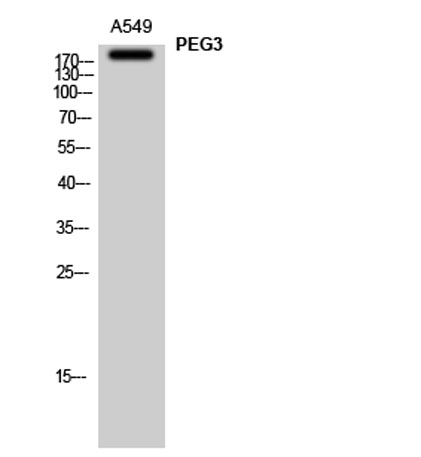
| Observed band (KD) | 181kD |
| Background | In human, ZIM2 and PEG3 are treated as two distinct genes though they share multiple 5' exons and a common promoter and both genes are paternally expressed (PMID:15203203). Alternative splicing events connect their shared 5' exons either with the remaining 4 exons unique to ZIM2, or with the remaining 2 exons unique to PEG3. In contrast, in other mammals ZIM2 does not undergo imprinting and, in mouse, cow, and likely other mammals as well, the ZIM2 and PEG3 genes do not share exons. Human PEG3 protein belongs to the Kruppel C2H2-type zinc finger protein family. PEG3 may play a role in cell proliferation and p53-mediated apoptosis. PEG3 has also shown tumor suppressor activity and tumorigenesis in glioma and ovarian cells. Alternative splicing of this PEG3 gene results in multiple transcript variants encoding distinct isoforms. [provided by RefSeq, Sep 2009], |
| Function | function:Induces apoptosis in cooperation with SIAH1A. Acts as a mediator between TP53/p53 and BAX in a neuronal death pathway that is activated by DNA damage. Acts synergistically with TRAF2 and inhibits TNF induced apoptosis through activation of NF-kappa-B (By similarity). Possesses a tumor suppressing activity in glioma cells.,function:May be involved in transcriptional regulation.,similarity:Belongs to the krueppel C2H2-type zinc-finger protein family.,similarity:Contains 1 KRAB domain.,similarity:Contains 1 SCAN box domain.,similarity:Contains 12 C2H2-type zinc fingers.,similarity:Contains 5 C2H2-type zinc fingers.,subunit:Homodimer. Interacts with SIAH1A and SIAH2. Interacts with TRAF2.,tissue specificity:Brain, glial cells, astrocytes, embryo, placenta, testis, ovary and utreus. In the placenta it is found in the layer of villous cytotrophoblast cells while in the ovary it is fou |
| Subcellular location | Nucleus . Cytoplasm . |
| Expression | Brain, glial cells, astrocytes, embryo, placenta, testis, ovary and uterus. In the placenta it is found in the layer of villous cytotrophoblast cells while in the ovary it is found in the cells of the ovarian stroma including the thecal layers around the follicles. Expression is highly repressed in glioma cell lines. |

Use collapsible tabs for more detailed information that will help customers make a purchasing decision.
Ex: Shipping and return policies, size guides, and other common questions.