 Français
 English
PKD2 (phospho Ser812) rabbit pAb
AO-06-ES6597-100

| ELK.No | ES6597 |
| Product name | PKD2 (phospho Ser812) rabbit pAb |
| Reactivity | Human;Mouse;Rat |
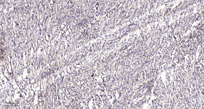
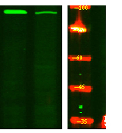
| Applications | WB;ELISA;IHC |
| Other name | PKD2; Polycystin-2; Autosomal dominant polycystic kidney disease type II protein; Polycystic kidney disease 2 protein; Polycystwin; R48321 |
| Size | 100μL |
| Unit price ($) | 248 |
| Human gene ID | 5311 |
| Human Swiss-Prot | Q13563 |
| Source | Rabbit |
| Isotype | IgG |
| Target | PKD2 |
| Fields | |
| Gene name | PKD2 |
| Protein name | Polycystin-2 |
| Human gene link | View Human Gene |
| Human Swiss link | View Human Swiss-Prot |
| Mouse gene ID | 18764 |
| Mouse gene link | View Mouse Gene |
| Mouse Swiss-Prot | O35245 |
| Mouse Swiss link | View Mouse Swiss-Prot |
| Rat gene ID | |
| Rat gene link | |
| Rat Swiss-Prot | |
| Rat Swiss link | |
| Immunogen | The antiserum was produced against synthesized peptide derived from human PKD2 around the phosphorylation site of Ser812. AA range:778-827 |
| Specificity | Phospho-PKD2 (S812) Polyclonal Antibody detects endogenous levels of PKD2S812. |
| Formulation | Liquid in PBS containing 50% glycerol, 0.5% BSA and 0.02% sodium azide. |
| Clonality | Polyclonal |
| Dilution | WB 1:500-2000;IHC-p 1:50-300; ELISA 2000-20000 |
| Purification | The antibody was affinity-purified from rabbit antiserum by affinity-chromatography using epitope-specific immunogen. |
| Concentration | 1 mg/ml |
| Storage stability | -20°C/1 year |
| Molecular Weight (Da) | 97kD |
| Observed band (KD) | |
| Background | polycystin 2, transient receptor potential cation channel(PKD2) Homo sapiens This gene encodes a member of the polycystin protein family. The encoded protein is a multi-pass membrane protein that functions as a calcium permeable cation channel, and is involved in calcium transport and calcium signaling in renal epithelial cells. This protein interacts with polycystin 1, and they may be partners in a common signaling cascade involved in tubular morphogenesis. Mutations in this gene are associated with autosomal dominant polycystic kidney disease type 2. [provided by RefSeq, Mar 2011], |
| Function | disease:Defects in PKD2 are the cause of polycystic kidney disease autosomal dominant type 2 (ADPKD2) [MIM:173900]. ADPKD2 represents approximately 15% of the cases of ADPKD, a common genetic disease affecting about 1:400 to 1:1000 individuals. ADPKD is characterized by progressive formation and enlargement of cysts in both kidneys, typically leading to end-stage renal disease in adult life. Cysts also occurs in the liver and other organs. ADPKD2 is clinically milder than ADPKD1 but it has a deleterious impact on overall life expectancy.,domain:The C-terminal coiled-coil domain binds calcium and undergoes a calcium-induced conformation change. It is implicated in oligomerization and the interaction with PKD1.,function:Functions as a calcium permeable cation channel. PKD1 and PKD2 may function through a common signaling pathway that is necessary for normal tubulogenesis.,online informatio |
| Subcellular location | Cell projection, cilium membrane ; Multi-pass membrane protein . Endoplasmic reticulum membrane ; Multi-pass membrane protein . Cell membrane ; Multi-pass membrane protein . Basolateral cell membrane . Cytoplasmic vesicle membrane . Golgi apparatus . PKD2 localization to the plasma and ciliary membranes requires PKD1. PKD1:PKD2 interaction is required to reach the Golgi apparatus form endoplasmic reticulum and then traffic to the cilia (By similarity). Retained in the endoplasmic reticulum by interaction with PACS1 and PACS2 (PubMed:15692563). Detected on kidney tubule basolateral membranes and basal cytoplasmic vesicles (PubMed:10770959). Cell surface and cilium localization requires GANAB (PubMed:27259053). . |
| Expression | Detected in fetal and adult kidney (PubMed:10770959). Detected at the thick ascending limb of the loop of Henle, at distal tubules, including the distal convoluted tubule and cortical collecting tubules, with weak staining of the collecting duct (PubMed:10770959). Detected on placenta syncytiotrophoblasts (at protein level) (PubMed:26269590). Strongly expressed in ovary, fetal and adult kidney, testis, and small intestine. Not detected in peripheral leukocytes. |

Use collapsible tabs for more detailed information that will help customers make a purchasing decision.
Ex: Shipping and return policies, size guides, and other common questions.