 Français
 English
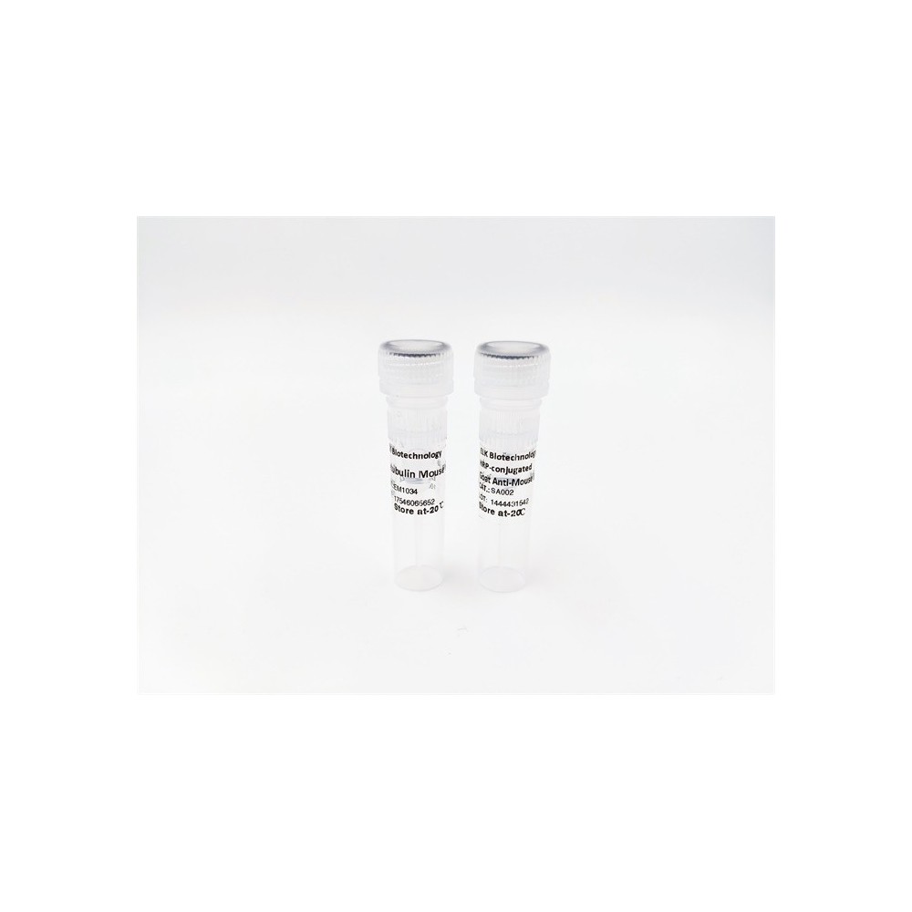

| ELK.No | ES6660 |
| Product name | RIAM rabbit pAb |
| Reactivity | Human;Rat;Mouse; |
| Applications | WB;IHC |
| Other name | APBB1IP; PREL1; RARP1; RIAM; Amyloid beta A4 precursor protein-binding family B member 1-interacting protein; APBB1-interacting protein 1; Proline-rich EVH1 ligand 1; PREL-1; Proline-rich protein 73; Rap1-GTP-interacting adapter molecule; R |
| Size | 50μL |
| Unit price ($) | 148 |
| Human gene ID | 54518 |
| Human Swiss-Prot | Q7Z5R6 |
| Source | Rabbit |
| Isotype | IgG |
| Target | RIAM |
| Fields | >>Rap1 signaling pathway;>>Platelet activation |
| Gene name | APBB1IP |
| Protein name | Amyloid beta A4 precursor protein-binding family B member 1-interacting protein |
| Human gene link | View Human Gene |
| Human Swiss link | View Human Swiss-Prot |
| Mouse gene ID | |
| Mouse gene link | |
| Mouse Swiss-Prot | Q8R5A3 |
| Mouse Swiss link | View Mouse Swiss-Prot |
| Rat gene ID | |
| Rat gene link | |
| Rat Swiss-Prot | |
| Rat Swiss link | |
| Immunogen | Synthesized peptide derived from RIAM . at AA range: 430-510 |
| Specificity | RIAM Polyclonal Antibody detects endogenous levels of RIAM protein. |
| Formulation | Liquid in PBS containing 50% glycerol, 0.5% BSA and 0.02% sodium azide. |
| Clonality | Polyclonal |
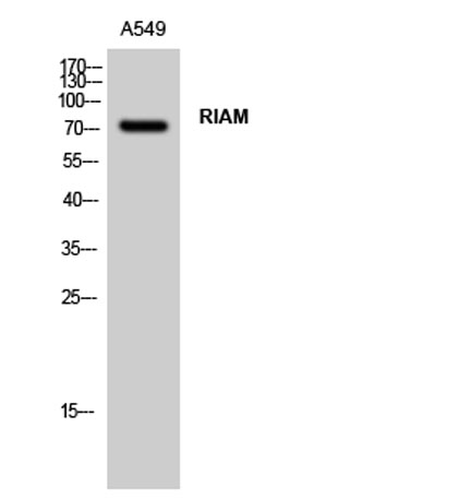
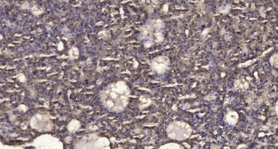
| Dilution | WB 1:500-2000;IHC-p 1:50-300 |
| Purification | The antibody was affinity-purified from rabbit antiserum by affinity-chromatography using epitope-specific immunogen. |
| Concentration | 1 mg/ml |
| Storage stability | -20°C/1 year |
| Molecular Weight (Da) | |
| Observed band (KD) | 73kD |
| Background | domain:The two Pro-rich regions are required for the suppression of AP1 transcription activity.,function:Appears to function in the signal transduction from Ras activation to actin cytoskeletal remodeling. Suppresses insulin-induced promoter activities through AP1 and SRE. Mediates Rap1-induced adhesion.,induction:Induced by all-trans-retinoic acid.,similarity:Belongs to the MRL family.,similarity:Contains 1 PH domain.,similarity:Contains 1 Ras-associating domain.,subcellular location:Colocalizes with ENA/VASP proteins at lamellipodia tips and focal adhesions, and F-actin at the leading edge. At the membrane surface, associates, via the PH domain, preferentially with the inositol phosphates, PtdIns(5)P and PtdIns(3)P. This binding appears to be necessary for the efficient interaction of the RA domain to Ras-GTPases.,subunit:Interacts, through the N-terminal Pro-rich region, with the WW domain of APBB1. Interacts with RAP1A, PFN1, VASP and ENAH.,tissue specificity:Widely expressed with high expression in thymus, spleen, lymph node, bone marrow and peripheral leukocytes., |
| Function | domain:The two Pro-rich regions are required for the suppression of AP1 transcription activity.,function:Appears to function in the signal transduction from Ras activation to actin cytoskeletal remodeling. Suppresses insulin-induced promoter activities through AP1 and SRE. Mediates Rap1-induced adhesion.,induction:Induced by all-trans-retinoic acid.,similarity:Belongs to the MRL family.,similarity:Contains 1 PH domain.,similarity:Contains 1 Ras-associating domain.,subcellular location:Colocalizes with ENA/VASP proteins at lamellipodia tips and focal adhesions, and F-actin at the leading edge. At the membrane surface, associates, via the PH domain, preferentially with the inositol phosphates, PtdIns(5)P and PtdIns(3)P. This binding appears to be necessary for the efficient interaction of the RA domain to Ras-GTPases.,subunit:Interacts, through the N-terminal Pro-rich region, with the WW d |
| Subcellular location | Cell membrane ; Peripheral membrane protein . Cell projection, lamellipodium . Cell junction, focal adhesion . Cytoplasm, cytoskeleton . Colocalizes with ENA/VASP proteins at lamellipodia tips and focal adhesions, and F-actin at the leading edge. At the membrane surface, associates, via the PH domain, preferentially with the inositol phosphates, PtdIns(5)P and PtdIns(3)P. This binding appears to be necessary for the efficient interaction of the RA domain to Ras-GTPases (By similarity). . |
| Expression | Widely expressed with high expression in thymus, spleen, lymph node, bone marrow and peripheral leukocytes. |

Use collapsible tabs for more detailed information that will help customers make a purchasing decision.
Ex: Shipping and return policies, size guides, and other common questions.