 Français
 English
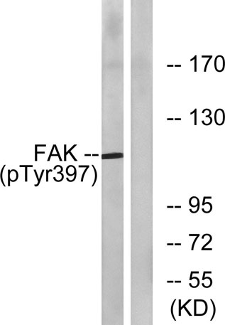
FAK (phospho Tyr397) rabbit pAb
AO-06-ES6902-100

| ELK.No | ES6902 |
| Product name | FAK (phospho Tyr397) rabbit pAb |
| Reactivity | Human;Mouse;Rat |
| Applications | WB;ELISA |
| Other name | PTK2; FAK; FAK1; Focal adhesion kinase 1; FADK 1; Focal adhesion kinase-related nonkinase; FRNK; Protein phosphatase 1 regulatory subunit 71; PPP1R71; Protein-tyrosine kinase 2; p125FAK; pp125FAK |
| Size | 100μL |
| Unit price ($) | 248 |
| Human gene ID | 5747 |
| Human Swiss-Prot | Q05397 |
| Source | Rabbit |
| Isotype | IgG |
| Target | FAK |
| Fields | >>Endocrine resistance;>>ErbB signaling pathway;>>Chemokine signaling pathway;>>PI3K-Akt signaling pathway;>>Axon guidance;>>VEGF signaling pathway;>>Focal adhesion;>>Leukocyte transendothelial migration;>>Regulation of actin cytoskeleton;>>Growth hormone synthesis, secretion and action;>>Bacterial invasion of epithelial cells;>>Shigellosis;>>Yersinia infection;>>Amoebiasis;>>Human cytomegalovirus infection;>>Human papillomavirus infection;>>Human immunodeficiency virus 1 infection;>>Pathways in cancer;>>Transcriptional misregulation in cancer;>>Proteoglycans in cancer;>>Chemical carcinogenesis - reactive oxygen species;>>Small cell lung cancer;>>Lipid and atherosclerosis;>>Fluid shear stress and atherosclerosis |
| Gene name | PTK2 |
| Protein name | Focal adhesion kinase 1 |
| Human gene link | View Human Gene |
| Human Swiss link | View Human Swiss-Prot |
| Mouse gene ID | 14083 |
| Mouse gene link | View Mouse Gene |
| Mouse Swiss-Prot | P34152 |
| Mouse Swiss link | View Mouse Swiss-Prot |
| Rat gene ID | 25614 |
| Rat gene link | View Rat Gene |
| Rat Swiss-Prot | O35346 |
| Rat Swiss link | View Rat Swiss-Prot |
| Immunogen | The antiserum was produced against synthesized peptide derived from human FAK around the phosphorylation site of Tyr397. AA range:364-413 |
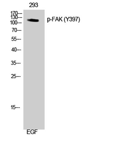
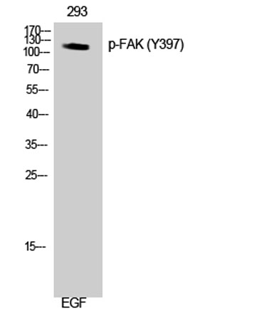
| Specificity | Phospho-FAK (Y397) Polyclonal Antibody detects endogenous levels of FAK protein only when phosphorylated at Y397. |
| Formulation | Liquid in PBS containing 50% glycerol, 0.5% BSA and 0.02% sodium azide. |
| Clonality | Polyclonal |
| Dilution | Western Blot: 1/500 - 1/2000. ELISA: 1/5000. Not yet tested in other applications. |
| Purification | The antibody was affinity-purified from rabbit antiserum by affinity-chromatography using epitope-specific immunogen. |
| Concentration | 1 mg/ml |
| Storage stability | -20°C/1 year |
| Molecular Weight (Da) | |
| Observed band (KD) | 119kD |
| Background | protein tyrosine kinase 2(PTK2) Homo sapiens This gene encodes a cytoplasmic protein tyrosine kinase which is found concentrated in the focal adhesions that form between cells growing in the presence of extracellular matrix constituents. The encoded protein is a member of the FAK subfamily of protein tyrosine kinases but lacks significant sequence similarity to kinases from other subfamilies. Activation of this gene may be an important early step in cell growth and intracellular signal transduction pathways triggered in response to certain neural peptides or to cell interactions with the extracellular matrix. Several transcript variants encoding different isoforms have been found for this gene, but the full-length natures of only four of them have been determined. [provided by RefSeq, Oct 2015], |
| Function | catalytic activity:ATP + a [protein]-L-tyrosine = ADP + a [protein]-L-tyrosine phosphate.,domain:The carboxy-terminal region is the site of focal adhesion targeting (FAT) sequence which mediates the localization of FAK1 to focal adhesions.,domain:The first Pro-rich domain interacts with the SH3 domain of CRK-associated substrate (BCAR1) and CASL.,function:Non-receptor protein-tyrosine kinase implicated in signaling pathways involved in cell motility, proliferation and apoptosis. Activated by tyrosine-phosphorylation in response to either integrin clustering induced by cell adhesion or antibody cross-linking, or via G-protein coupled receptor (GPCR) occupancy by ligands such as bombesin or lysophosphatidic acid, or via LDL receptor occupancy. Plays a potential role in oncogenic transformations resulting in increased kinase activity.,PTM:Phosphorylated on 6 tyrosine residues upon activatio |
| Subcellular location | Cell junction, focal adhesion. Cell membrane; Peripheral membrane protein; Cytoplasmic side. Cytoplasm, cell cortex. Cytoplasm, cytoskeleton. Cytoplasm, cytoskeleton, microtubule organizing center, centrosome . Nucleus. Cytoplasm, cytoskeleton, cilium basal body . Constituent of focal adhesions. Detected at microtubules. |
| Expression | Detected in B and T-lymphocytes. Isoform 1 and isoform 6 are detected in lung fibroblasts (at protein level). Ubiquitous. Expressed in epithelial cells (at protein level) (PubMed:31630787). |

Use collapsible tabs for more detailed information that will help customers make a purchasing decision.
Ex: Shipping and return policies, size guides, and other common questions.