 Français
 English
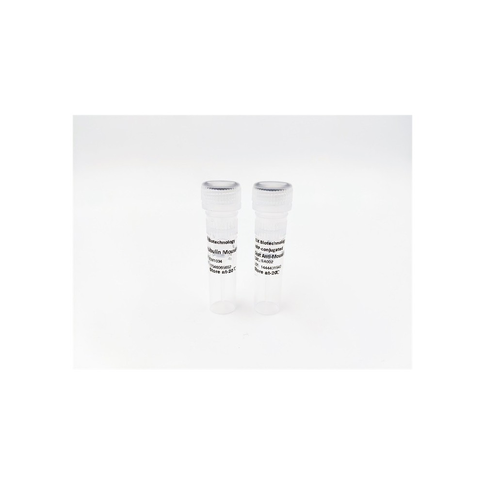
SH-PTP1 (phospho Tyr564) rabbit pAb
AO-06-ES6931-50

| ELK.No | ES6931 |
| Product name | SH-PTP1 (phospho Tyr564) rabbit pAb |
| Reactivity | Human;Monkey |
| Applications | WB;ELISA;IHC |
| Other name | PTPN6; HCP; PTP1C; Tyrosine-protein phosphatase non-receptor type 6; Hematopoietic cell protein-tyrosine phosphatase; Protein-tyrosine phosphatase 1C; PTP-1C; Protein-tyrosine phosphatase SHP-1; SH-PTP1 |
| Size | 50μL |
| Unit price ($) | 148 |
| Human gene ID | 5777 |
| Human Swiss-Prot | P29350 |
| Source | Rabbit |
| Isotype | IgG |
| Target | SH-PTP1 |
| Fields | >>Adherens junction;>>JAK-STAT signaling pathway;>>Natural killer cell mediated cytotoxicity;>>T cell receptor signaling pathway;>>B cell receptor signaling pathway;>>Pathogenic Escherichia coli infection;>>Leishmaniasis;>>Proteoglycans in cancer;>>PD-L1 expression and PD-1 checkpoint pathway in cancer |
| Gene name | PTPN6 |
| Protein name | Tyrosine-protein phosphatase non-receptor type 6 |
| Human gene link | View Human Gene |
| Human Swiss link | View Human Swiss-Prot |
| Mouse gene ID | |
| Mouse gene link | |
| Mouse Swiss-Prot | P29351 |
| Mouse Swiss link | View Mouse Swiss-Prot |
| Rat gene ID | |
| Rat gene link | |
| Rat Swiss-Prot | |
| Rat Swiss link | |
| Immunogen | The antiserum was produced against synthesized peptide derived from human SHP-1 around the phosphorylation site of Tyr564. AA range:530-579 |
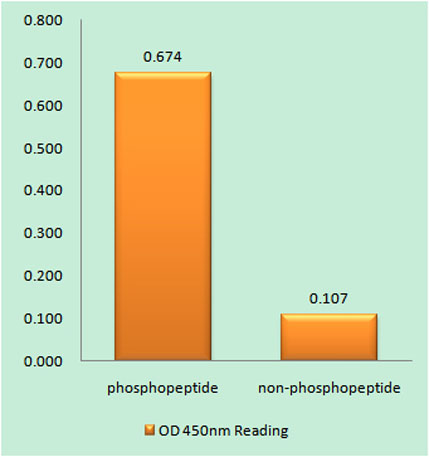
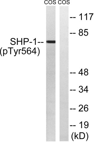
| Specificity | Phospho-SH-PTP1 (Y564) Polyclonal Antibody detects endogenous levels of SH-PTP1 protein only when phosphorylated at Y564. |
| Formulation | Liquid in PBS containing 50% glycerol, 0.5% BSA and 0.02% sodium azide. |
| Clonality | Polyclonal |
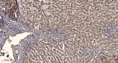
| Dilution | WB 1:500-2000;IHC-p 1:50-300; ELISA 2000-20000 |
| Purification | The antibody was affinity-purified from rabbit antiserum by affinity-chromatography using epitope-specific immunogen. |
| Concentration | 1 mg/ml |
| Storage stability | -20°C/1 year |
| Molecular Weight (Da) | |
| Observed band (KD) | 70kD |
| Background | The protein encoded by this gene is a member of the protein tyrosine phosphatase (PTP) family. PTPs are known to be signaling molecules that regulate a variety of cellular processes including cell growth, differentiation, mitotic cycle, and oncogenic transformation. N-terminal part of this PTP contains two tandem Src homolog (SH2) domains, which act as protein phospho-tyrosine binding domains, and mediate the interaction of this PTP with its substrates. This PTP is expressed primarily in hematopoietic cells, and functions as an important regulator of multiple signaling pathways in hematopoietic cells. This PTP has been shown to interact with, and dephosphorylate a wide spectrum of phospho-proteins involved in hematopoietic cell signaling. Multiple alternatively spliced variants of this gene, which encode distinct isoforms, have been reported. [provided by RefSeq, Jul |
| Function | catalytic activity:Protein tyrosine phosphate + H(2)O = protein tyrosine + phosphate.,function:Plays a key role in hematopoiesis. This PTPase activity may directly link growth factor receptors and other signaling proteins through protein-tyrosine phosphorylation. The SH2 regions may interact with other cellular components to modulate its own phosphatase activity against interacting substrates. Together with MTUS1, induces UBE2V2 expression upon angiotensin II stimulation.,PTM:Phosphorylated on serine and tyrosine residues.,similarity:Belongs to the protein-tyrosine phosphatase family. Non-receptor class 2 subfamily.,similarity:Contains 1 tyrosine-protein phosphatase domain.,similarity:Contains 2 SH2 domains.,subcellular location:In neurons, translocates into the nucleus after treatment with angiotensin II.,subunit:Monomer. Interacts with MTUS1 (By similarity). Binds PTPNS1, LILRB1 and LI |
| Subcellular location | Cytoplasm. Nucleus. In neurons, translocates into the nucleus after treatment with angiotensin II (By similarity). Shuttles between the cytoplasm and nucleus via its association with PDPK1. . |
| Expression | Isoform 1 is expressed in hematopoietic cells. Isoform 2 is expressed in non-hematopoietic cells. |

Use collapsible tabs for more detailed information that will help customers make a purchasing decision.
Ex: Shipping and return policies, size guides, and other common questions.