 Français
 English

| ELK.No | ES6942 |
| Product name | CD45 rabbit pAb |
| Reactivity | Human;Mouse;Rat |
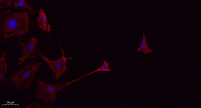
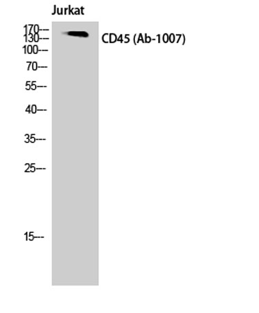
| Applications | WB;IHC;IF;ELISA |
| Other name | PTPRC; CD45; Receptor-type tyrosine-protein phosphatase C; Leukocyte common antigen; L-CA; T200; CD antigen CD45 |
| Size | 50μL |
| Unit price ($) | 148 |
| Human gene ID | 5788 |
| Human Swiss-Prot | P08575 |
| Source | Rabbit |
| Isotype | IgG |
| Target | CD45 |
| Fields | >>Cell adhesion molecules;>>T cell receptor signaling pathway;>>Fc gamma R-mediated phagocytosis;>>Salmonella infection;>>Primary immunodeficiency |
| Gene name | PTPRC |
| Protein name | Receptor-type tyrosine-protein phosphatase C |
| Human gene link | View Human Gene |
| Human Swiss link | View Human Swiss-Prot |
| Mouse gene ID | |
| Mouse gene link | |
| Mouse Swiss-Prot | P06800 |
| Mouse Swiss link | View Mouse Swiss-Prot |
| Rat gene ID | 24699 |
| Rat gene link | View Rat Gene |
| Rat Swiss-Prot | P04157 |
| Rat Swiss link | View Rat Swiss-Prot |
| Immunogen | The antiserum was produced against synthesized peptide derived from human CD45. AA range:981-1030 |
| Specificity | CD45 Polyclonal Antibody detects endogenous levels of CD45 protein. |
| Formulation | Liquid in PBS containing 50% glycerol, 0.5% BSA and 0.02% sodium azide. |
| Clonality | Polyclonal |
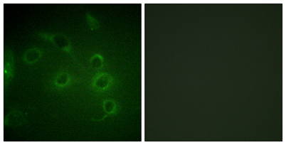
| Dilution | Western Blot: 1/500 - 1/2000. IHC-p: 1:100-300 ELISA: 1/20000. IF 1:100-300 Not yet tested in other applications. |
| Purification | The antibody was affinity-purified from rabbit antiserum by affinity-chromatography using epitope-specific immunogen. |
| Concentration | 1 mg/ml |
| Storage stability | -20°C/1 year |
| Molecular Weight (Da) | 147kD |
| Observed band (KD) | |
| Background | The protein encoded by this gene is a member of the protein tyrosine phosphatase (PTP) family. PTPs are known to be signaling molecules that regulate a variety of cellular processes including cell growth, differentiation, mitosis, and oncogenic transformation. This PTP contains an extracellular domain, a single transmembrane segment and two tandem intracytoplasmic catalytic domains, and thus is classified as a receptor type PTP. This PTP has been shown to be an essential regulator of T- and B-cell antigen receptor signaling. It functions through either direct interaction with components of the antigen receptor complexes, or by activating various Src family kinases required for the antigen receptor signaling. This PTP also suppresses JAK kinases, and thus functions as a regulator of cytokine receptor signaling. Alternatively spliced transcripts variants of this gene, which enc |
| Function | alternative products:At least 8 isoforms are produced,catalytic activity:Protein tyrosine phosphate + H(2)O = protein tyrosine + phosphate.,disease:Defects in PTPRC are a cause of severe combined immunodeficiency autosomal recessive T-cell-negative/B-cell-positive/NK-cell-positive (T(-)B(+)NK(+)SCID) [MIM:608971]. SCID refers to a genetically and clinically heterogeneous group of rare congenital disorders characterized by impairment of both humoral and cell-mediated immunity, leukopenia, and low or absent antibody levels. Patients with SCID present in infancy with recurrent, persistent infections by opportunistic organisms. The common characteristic of all types of SCID is absence of T-cell-mediated cellular immunity due to a defect in T-cell development.,disease:Genetic variations in PTPRC are involved in multiple sclerosis susceptibility (MS) [MIM:126200]. MS is a neurodegenerative dis |
| Subcellular location | Cell membrane ; Single-pass type I membrane protein . Membrane raft . Colocalized with DPP4 in membrane rafts. . |
| Expression | Isoform 1: Detected in thymocytes. Isoform 2: Detected in thymocytes. Isoform 3: Detected in thymocytes. Isoform 4: Not detected in thymocytes. Isoform 5: Detected in thymocytes. Isoform 6: Not detected in thymocytes. Isoform 7: Detected in thymocytes. Isoform 8: Not detected in thymocytes. |

Use collapsible tabs for more detailed information that will help customers make a purchasing decision.
Ex: Shipping and return policies, size guides, and other common questions.