 Français
 English
Ret (phospho Tyr1015) rabbit pAb
AO-06-ES7031-100

| ELK.No | ES7031 |
| Product name | Ret (phospho Tyr1015) rabbit pAb |
| Reactivity | Human;Mouse;Rat;Monkey |
| Applications | WB;IHC |
| Other name | RET; CDHF12; CDHR16; PTC; RET51; Proto-oncogene tyrosine-protein kinase receptor Ret; Cadherin family member 12; Proto-oncogene c-Ret |
| Size | 100μL |
| Unit price ($) | 248 |
| Human gene ID | 5979 |
| Human Swiss-Prot | P07949 |
| Source | Rabbit |
| Isotype | IgG |
| Target | Ret |
| Fields | >>Calcium signaling pathway;>>Pathways in cancer;>>Thyroid cancer;>>Non-small cell lung cancer;>>Central carbon metabolism in cancer |
| Gene name | RET |
| Protein name | Proto-oncogene tyrosine-protein kinase receptor Ret |
| Human gene link | View Human Gene |
| Human Swiss link | View Human Swiss-Prot |
| Mouse gene ID | 19713 |
| Mouse gene link | View Mouse Gene |
| Mouse Swiss-Prot | P35546 |
| Mouse Swiss link | View Mouse Swiss-Prot |
| Rat gene ID | |
| Rat gene link | |
| Rat Swiss-Prot | |
| Rat Swiss link | |
| Immunogen | The antiserum was produced against synthesized peptide derived from human Ret around the phosphorylation site of Tyr1015. AA range:991-1040 |
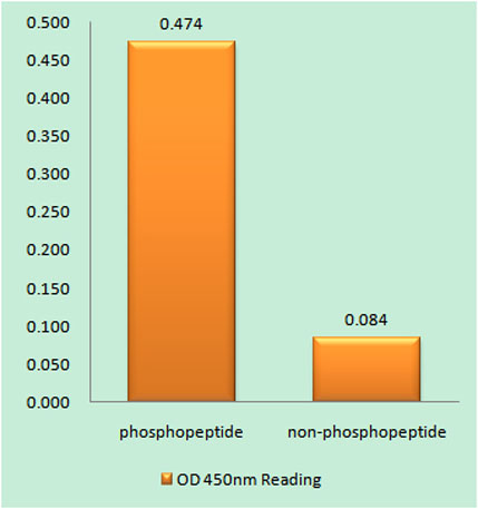
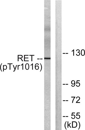
| Specificity | Phospho-Ret (Y1015) Polyclonal Antibody detects endogenous levels of Ret protein only when phosphorylated at Y1015. |
| Formulation | Liquid in PBS containing 50% glycerol, 0.5% BSA and 0.02% sodium azide. |
| Clonality | Polyclonal |
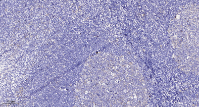
| Dilution | WB 1:500-2000;IHC-p 1:50-300 |
| Purification | The antibody was affinity-purified from rabbit antiserum by affinity-chromatography using epitope-specific immunogen. |
| Concentration | 1 mg/ml |
| Storage stability | -20°C/1 year |
| Molecular Weight (Da) | |
| Observed band (KD) | 124kD |
| Background | ret proto-oncogene(RET) Homo sapiens This gene, a member of the cadherin superfamily, encodes one of the receptor tyrosine kinases, which are cell-surface molecules that transduce signals for cell growth and differentiation. This gene plays a crucial role in neural crest development, and it can undergo oncogenic activation in vivo and in vitro by cytogenetic rearrangement. Mutations in this gene are associated with the disorders multiple endocrine neoplasia, type IIA, multiple endocrine neoplasia, type IIB, Hirschsprung disease, and medullary thyroid carcinoma. Two transcript variants encoding different isoforms have been found for this gene. Additional transcript variants have been described but their biological validity has not been confirmed. [provided by RefSeq, Jul 2008], |
| Function | catalytic activity:ATP + a [protein]-L-tyrosine = ADP + a [protein]-L-tyrosine phosphate.,disease:Chromosomal aberrations involving RET are a cause of thyroid papillary carcinoma (PACT) [MIM:188550]. Inversion inv(10)(q11.2;q21) generates the RET/CCDC6 (PTC1) oncogene; inversion inv(10)(q11.2;q11.2) generates the RET/NCOA4 (PTC3) oncogene; translocation t(10;14)(q11;q32) with GOLGA5 generates the RET/GOLGA5 (PTC5) oncogene; translocation t(8;10)(p21.3;q11.2) with PCM1 generates the PCM1/RET fusion; translocation t(6;10)(p21.3;q11.2) with RFP generates the Delta RFP/RET oncogene; translocation t(1;10)(p13;q11) with TRIM33 generates the TRIM33/RET (PTC7) oncogene; translocation t(7;10)(q32;q11) with TIF1 generates the TIF1/RET (PTC6) oncogene. The PTC5 oncogene has been found in 2 cases of PACT in children exposed to radioactive fallout after Chernobyl.,disease:Defects in RET are a cause o |
| Subcellular location | Cell membrane ; Single-pass type I membrane protein . Endosome membrane ; Single-pass type I membrane protein . Predominantly located on the plasma membrane. In the presence of SORL1 and GFRA1, directed to endosomes. . |
| Expression | Blood,Brain,Fibroblast,Leukocyte,Neural crest,Peripheral blood leukocyte,Thyroid papillary |

Use collapsible tabs for more detailed information that will help customers make a purchasing decision.
Ex: Shipping and return policies, size guides, and other common questions.