 Français
 English
Ataxin-1 (phospho Ser776) rabbit pAb
AO-06-ES7119-100

| ELK.No | ES7119 |
| Product name | Ataxin-1 (phospho Ser776) rabbit pAb |
| Reactivity | Human;Mouse |
| Applications | WB;IHC;IF;ELISA |
| Other name | ATXN1; ATX1; SCA1; Ataxin-1; Spinocerebellar ataxia type 1 protein |
| Size | 100μL |
| Unit price ($) | 248 |
| Human gene ID | 6310 |
| Human Swiss-Prot | P54253 |
| Source | Rabbit |
| Isotype | IgG |
| Target | Ataxin-1 |
| Fields | >>Notch signaling pathway;>>Spinocerebellar ataxia;>>Pathways of neurodegeneration - multiple diseases |
| Gene name | ATXN1 |
| Protein name | Ataxin-1 |
| Human gene link | View Human Gene |
| Human Swiss link | View Human Swiss-Prot |
| Mouse gene ID | 20238 |
| Mouse gene link | View Mouse Gene |
| Mouse Swiss-Prot | P54254 |
| Mouse Swiss link | View Mouse Swiss-Prot |
| Rat gene ID | |
| Rat gene link | |
| Rat Swiss-Prot | |
| Rat Swiss link | |
| Immunogen | The antiserum was produced against synthesized peptide derived from human Ataxin 1 around the phosphorylation site of Ser776. AA range:742-791 |
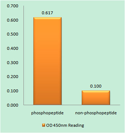
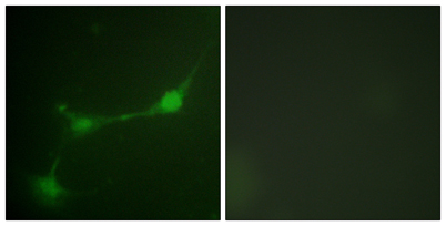
| Specificity | Phospho-Ataxin-1 (S776) Polyclonal Antibody detects endogenous levels of Ataxin-1 protein only when phosphorylated at S776. |
| Formulation | Liquid in PBS containing 50% glycerol, 0.5% BSA and 0.02% sodium azide. |
| Clonality | Polyclonal |
| Dilution | Western Blot: 1/500 - 1/2000. Immunohistochemistry: 1/100 - 1/300. Immunofluorescence: 1/200 - 1/1000. ELISA: 1/10000. Not yet tested in other applications. |
| Purification | The antibody was affinity-purified from rabbit antiserum by affinity-chromatography using epitope-specific immunogen. |
| Concentration | 1 mg/ml |
| Storage stability | -20°C/1 year |
| Molecular Weight (Da) | |
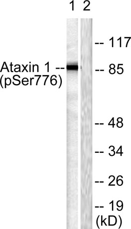
| Observed band (KD) | 87kD |
| Background | ataxin 1(ATXN1) Homo sapiens The autosomal dominant cerebellar ataxias (ADCA) are a heterogeneous group of neurodegenerative disorders characterized by progressive degeneration of the cerebellum, brain stem and spinal cord. Clinically, ADCA has been divided into three groups: ADCA types I-III. ADCAI is genetically heterogeneous, with five genetic loci, designated spinocerebellar ataxia (SCA) 1, 2, 3, 4 and 6, being assigned to five different chromosomes. ADCAII, which always presents with retinal degeneration (SCA7), and ADCAIII often referred to as the `pure' cerebellar syndrome (SCA5), are most likely homogeneous disorders. Several SCA genes have been cloned and shown to contain CAG repeats in their coding regions. ADCA is caused by the expansion of the CAG repeats, producing an elongated polyglutamine tract in the corresponding protein. The expanded repeats are variable in size and unstable, usually increasing in size when transmitted |
| Function | alternative products:At least 2 isoforms are produced,disease:Defects in ATXN1 are the cause of spinocerebellar ataxia type 1 (SCA1) [MIM:164400]; also known as olivopontocerebellar atrophy I (OPCA I or OPCA1). Spinocerebellar ataxia is a clinically and genetically heterogeneous group of cerebellar disorders. Patients show progressive incoordination of gait and often poor coordination of hands, speech and eye movements, due to cerebellum degeneration with variable involvement of the brainstem and spinal cord. SCA1 belongs to the autosomal dominant cerebellar ataxias type I (ADCA I) which are characterized by cerebellar ataxia in combination with additional clinical features like optic atrophy, ophthalmoplegia, bulbar and extrapyramidal signs, peripheral neuropathy and dementia. SCA1 is caused by expansion of a CAG repeat in the coding region of ATXN1. Longer expansions result in earlier |
| Subcellular location | Cytoplasm . Nucleus . Colocalizes with USP7 in the nucleus. . |
| Expression | Widely expressed throughout the body. |

Use collapsible tabs for more detailed information that will help customers make a purchasing decision.
Ex: Shipping and return policies, size guides, and other common questions.