 Français
 English
Ezrin (phospho Tyr146) rabbit pAb
AO-06-ES7502-50

| ELK.No | ES7502 |
| Product name | Ezrin (phospho Tyr146) rabbit pAb |
| Reactivity | Human;Mouse;Rat |
| Applications | WB;ELISA |
| Other name | EZR; VIL2; Ezrin; Cytovillin; Villin-2; p81 |
| Size | 50μL |
| Unit price ($) | 148 |
| Human gene ID | 7430 |
| Human Swiss-Prot | P15311 |
| Source | Rabbit |
| Isotype | IgG |
| Target | Ezrin |
| Fields | >>Tight junction;>>Leukocyte transendothelial migration;>>Regulation of actin cytoskeleton;>>Gastric acid secretion;>>Pathogenic Escherichia coli infection;>>Proteoglycans in cancer;>>MicroRNAs in cancer |
| Gene name | EZR |
| Protein name | Ezrin |
| Human gene link | View Human Gene |
| Human Swiss link | View Human Swiss-Prot |
| Mouse gene ID | 22350 |
| Mouse gene link | View Mouse Gene |
| Mouse Swiss-Prot | P26040 |
| Mouse Swiss link | View Mouse Swiss-Prot |
| Rat gene ID | 54319 |
| Rat gene link | View Rat Gene |
| Rat Swiss-Prot | P31977 |
| Rat Swiss link | View Rat Swiss-Prot |
| Immunogen | The antiserum was produced against synthesized peptide derived from human Ezrin around the phosphorylation site of Tyr146. AA range:111-160 |
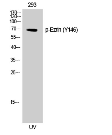
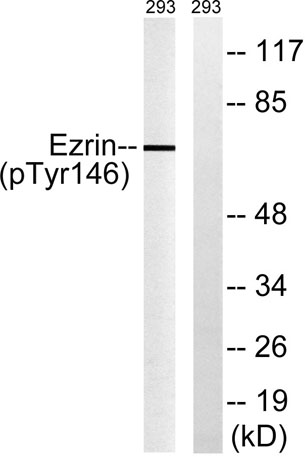
| Specificity | Phospho-Ezrin (Y146) Polyclonal Antibody detects endogenous levels of Ezrin protein only when phosphorylated at Y146. |
| Formulation | Liquid in PBS containing 50% glycerol, 0.5% BSA and 0.02% sodium azide. |
| Clonality | Polyclonal |
| Dilution | Western Blot: 1/500 - 1/2000. ELISA: 1/40000. Not yet tested in other applications. |
| Purification | The antibody was affinity-purified from rabbit antiserum by affinity-chromatography using epitope-specific immunogen. |
| Concentration | 1 mg/ml |
| Storage stability | -20°C/1 year |
| Molecular Weight (Da) | |
| Observed band (KD) | 69kD |
| Background | The cytoplasmic peripheral membrane protein encoded by this gene functions as a protein-tyrosine kinase substrate in microvilli. As a member of the ERM protein family, this protein serves as an intermediate between the plasma membrane and the actin cytoskeleton. This protein plays a key role in cell surface structure adhesion, migration and organization, and it has been implicated in various human cancers. A pseudogene located on chromosome 3 has been identified for this gene. Alternatively spliced variants have also been described for this gene. [provided by RefSeq, Jul 2008], |
| Function | developmental stage:Very strong staining is detected in the Purkinje cell layer and in part of the molecular layer of the infant brain compared to adult brain.,function:Probably involved in connections of major cytoskeletal structures to the plasma membrane. In epithelial cells, required for the formation of microvilli and membrane ruffles on the apical pole. Along with PLEKHG6, required for normal macropinocytosis.,PTM:Phosphorylated by tyrosine-protein kinases.,similarity:Contains 1 FERM domain.,subcellular location:Localization to the apical membrane of parietal cells depends on the interaction with MPP5. Localizes to cell extensions and peripheral processes of astrocytes (By similarity). Microvillar peripheral membrane protein (cytoplasmic side).,subunit:Interacts with MPP5 (By similarity). Interacts with SLC9A3R1 and SCYL3/PACE1. Interacts with PLEKHG6. Interacts with NGX6.,tissue s |
| Subcellular location | Apical cell membrane ; Peripheral membrane protein ; Cytoplasmic side . Cell projection . Cell projection, microvillus membrane ; Peripheral membrane protein ; Cytoplasmic side . Cell projection, ruffle membrane ; Peripheral membrane protein ; Cytoplasmic side . Cytoplasm, cell cortex . Cytoplasm, cytoskeleton . Cell projection, microvillus . Localization to the apical membrane of parietal cells depends on the interaction with PALS1. Localizes to cell extensions and peripheral processes of astrocytes (By similarity). Microvillar peripheral membrane protein (cytoplasmic side). . |
| Expression | Expressed in cerebral cortex, basal ganglia, hippocampus, hypophysis, and optic nerve. Weakly expressed in brain stem and diencephalon. Stronger expression was detected in gray matter of frontal lobe compared to white matter (at protein level). Component of the microvilli of intestinal epithelial cells. Preferentially expressed in astrocytes of hippocampus, frontal cortex, thalamus, parahippocampal cortex, amygdala, insula, and corpus callosum. Not detected in neurons in most tissues studied. |

Use collapsible tabs for more detailed information that will help customers make a purchasing decision.
Ex: Shipping and return policies, size guides, and other common questions.