 Français
 English
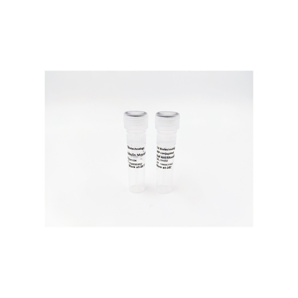

| ELK.No | ES7840 |
| Product name | p63 rabbit pAb |
| Reactivity | Human;Mouse;Rat;Monkey |
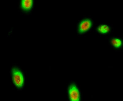
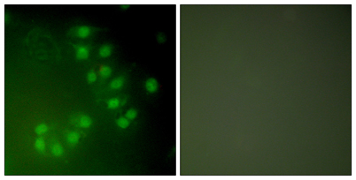
| Applications | WB;IHC;IF;ELISA |
| Other name | TP63; KET; P63; P73H; P73L; TP73L; Tumor protein 63; p63; Chronic ulcerative stomatitis protein; CUSP; Keratinocyte transcription factor KET; Transformation-related protein 63; TP63; Tumor protein p73-like; p73L; p40; p51 |
| Size | 100μL |
| Unit price ($) | 248 |
| Human gene ID | 8626 |
| Human Swiss-Prot | Q9H3D4 |
| Source | Rabbit |
| Isotype | IgG |
| Target | p40/p63 |
| Fields | >>MicroRNAs in cancer |
| Gene name | TP63 |
| Protein name | Tumor protein 63 |
| Human gene link | View Human Gene |
| Human Swiss link | View Human Swiss-Prot |
| Mouse gene ID | 22061 |
| Mouse gene link | View Mouse Gene |
| Mouse Swiss-Prot | O88898 |
| Mouse Swiss link | View Mouse Swiss-Prot |
| Rat gene ID | 246334 |
| Rat gene link | View Rat Gene |
| Rat Swiss-Prot | Q9JJP6 |
| Rat Swiss link | View Rat Swiss-Prot |
| Immunogen | The antiserum was produced against synthesized peptide derived from human p63. AA range:631-680 |
| Specificity | p63 Polyclonal Antibody detects endogenous levels of p63 protein. |
| Formulation | Liquid in PBS containing 50% glycerol, 0.5% BSA and 0.02% sodium azide. |
| Clonality | Polyclonal |
| Dilution | Western Blot: 1/500 - 1/2000. Immunohistochemistry: 1/100 - 1/300. Immunofluorescence: 1/200 - 1/1000. ELISA: 1/20000. Not yet tested in other applications. |
| Purification | The antibody was affinity-purified from rabbit antiserum by affinity-chromatography using epitope-specific immunogen. |
| Concentration | 1 mg/ml |
| Storage stability | -20°C/1 year |
| Molecular Weight (Da) | |
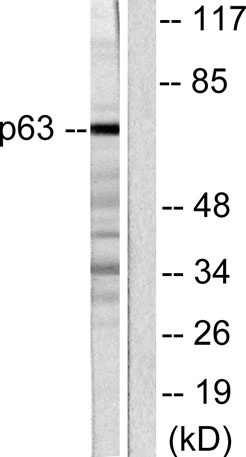
| Observed band (KD) | 77kD |
| Background | tumor protein p63(TP63) Homo sapiens This gene encodes a member of the p53 family of transcription factors. The functional domains of p53 family proteins include an N-terminal transactivation domain, a central DNA-binding domain and an oligomerization domain. Alternative splicing of this gene and the use of alternative promoters results in multiple transcript variants encoding different isoforms that vary in their functional properties. These isoforms function during skin development and maintenance, adult stem/progenitor cell regulation, heart development and premature aging. Some isoforms have been found to protect the germline by eliminating oocytes or testicular germ cells that have suffered DNA damage. Mutations in this gene are associated with ectodermal dysplasia, and cleft lip/palate syndrome 3 (EEC3); split-hand/foot malformation 4 (SHFM4); ankyloblepharon-ectodermal defects-cleft lip/palate; ADULT syndrome (acro-dermato-ungual-lacrim |
| Function | cofactor:Binds 1 zinc ion per subunit.,disease:Defects in TP63 are a cause of cervical, colon, head and neck, lung and ovarian cancers.,disease:Defects in TP63 are a cause of ectodermal dysplasia Rapp-Hodgkin type (EDRH) [MIM:129400]; also called Rapp-Hodgkin syndrome or anhidrotic ectodermal dysplasia with cleft lip/palate. Ectodermal dysplasia defines a heterogeneous group of disorders due to abnormal development of two or more ectodermal structures. EDRH is characterized by the combination of anhidrotic ectodermal dysplasia, cleft lip, and cleft palate. The clinical syndrome is comprised of a characteristic facies (narrow nose and small mouth), wiry, slow-growing, and uncombable hair, sparse eyelashes and eyebrows, obstructed lacrimal puncta/epiphora, bilateral stenosis of external auditory canals, microsomia, hypodontia, cone-shaped incisors, enamel hypoplasia, dystrophic nails, and |
| Subcellular location | Nucleus . |
| Expression | Widely expressed, notably in heart, kidney, placenta, prostate, skeletal muscle, testis and thymus, although the precise isoform varies according to tissue type. Progenitor cell layers of skin, breast, eye and prostate express high levels of DeltaN-type isoforms. Isoform 10 is predominantly expressed in skin squamous cell carcinomas, but not in normal skin tissues. |

Use collapsible tabs for more detailed information that will help customers make a purchasing decision.
Ex: Shipping and return policies, size guides, and other common questions.