 Français
 English
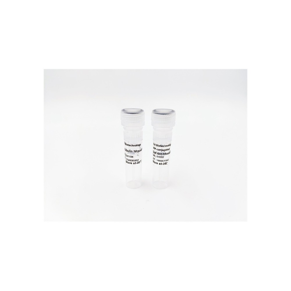
Cbl (phospho Tyr700) rabbit pAb
AO-06-ES7851-50

| ELK.No | ES7851 |
| Product name | Cbl (phospho Tyr700) rabbit pAb |
| Reactivity | Human;Mouse;Rat |
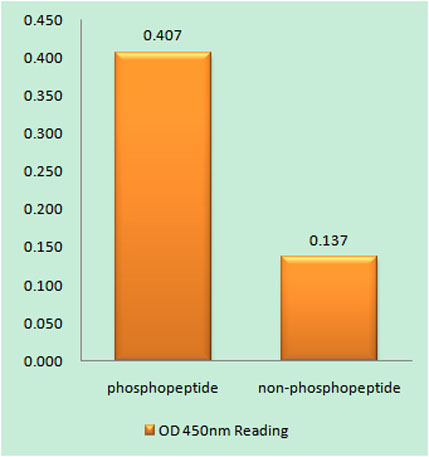
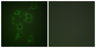
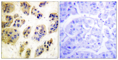
| Applications | WB;IHC;IF;ELISA |
| Other name | CBL; CBL2; RNF55; E3 ubiquitin-protein ligase CBL; Casitas B-lineage lymphoma proto-oncogene; Proto-oncogene c-Cbl; RING finger protein 55; Signal transduction protein CBL |
| Size | 50μL |
| Unit price ($) | 148 |
| Human gene ID | 867 |
| Human Swiss-Prot | P22681 |
| Source | Rabbit |
| Isotype | IgG |
| Target | Cbl |
| Fields | >>ErbB signaling pathway;>>Ubiquitin mediated proteolysis;>>Endocytosis;>>Insulin signaling pathway;>>Bacterial invasion of epithelial cells;>>Pathways in cancer;>>Proteoglycans in cancer;>>Chronic myeloid leukemia |
| Gene name | CBL |
| Protein name | E3 ubiquitin-protein ligase CBL |
| Human gene link | View Human Gene |
| Human Swiss link | View Human Swiss-Prot |
| Mouse gene ID | 12402 |
| Mouse gene link | View Mouse Gene |
| Mouse Swiss-Prot | P22682 |
| Mouse Swiss link | View Mouse Swiss-Prot |
| Rat gene ID | |
| Rat gene link | |
| Rat Swiss-Prot | |
| Rat Swiss link | |
| Immunogen | The antiserum was produced against synthesized peptide derived from human CBL around the phosphorylation site of Tyr700. AA range:666-715 |
| Specificity | Phospho-Cbl (Y700) Polyclonal Antibody detects endogenous levels of Cbl protein only when phosphorylated at Y700. |
| Formulation | Liquid in PBS containing 50% glycerol, 0.5% BSA and 0.02% sodium azide. |
| Clonality | Polyclonal |
| Dilution | Western Blot: 1/500 - 1/2000. Immunohistochemistry: 1/100 - 1/300. Immunofluorescence: 1/200 - 1/1000. ELISA: 1/10000. Not yet tested in other applications. |
| Purification | The antibody was affinity-purified from rabbit antiserum by affinity-chromatography using epitope-specific immunogen. |
| Concentration | 1 mg/ml |
| Storage stability | -20°C/1 year |
| Molecular Weight (Da) | |
| Observed band (KD) | 120kD |
| Background | Cbl proto-oncogene(CBL) Homo sapiens This gene is a proto-oncogene that encodes a RING finger E3 ubiquitin ligase. The encoded protein is one of the enzymes required for targeting substrates for degradation by the proteasome. This protein mediates the transfer of ubiquitin from ubiquitin conjugating enzymes (E2) to specific substrates. This protein also contains an N-terminal phosphotyrosine binding domain that allows it to interact with numerous tyrosine-phosphorylated substrates and target them for proteasome degradation. As such it functions as a negative regulator of many signal transduction pathways. This gene has been found to be mutated or translocated in many cancers including acute myeloid leukaemia, and expansion of CGG repeats in the 5' UTR has been associated with Jacobsen syndrome. Mutations in this gene are also the cause of Noonan syndrome-like disorder. [provided by RefSeq, Jul 2016], |
| Function | disease:Can be converted to an oncogenic protein by deletions or mutations that disturb its ability to down-regulate RTKs.,domain:The N-terminus is composed of the phosphotyrosine binding (PTB) domain, a short linker region and the RING-type zinc finger. The PTB domain, which is also called TKB (tyrosine kinase binding) domain, is composed of three different subdomains: a four-helix bundle (4H), a calcium-binding EF hand and a divergent SH2 domain.,domain:The RING-type zinc finger domain mediates binding to an E2 ubiquitin-conjugating enzyme.,function:Participates in signal transduction in hematopoietic cells. Adapter protein that functions as a negative regulator of many signaling pathways that start from receptors at the cell surface. Acts as an E3 ubiquitin-protein ligase, which accepts ubiquitin from specific E2 ubiquitin-conjugating enzymes, and then transfers it to substrates promo |
| Subcellular location | Cytoplasm. Cell membrane. Cell projection, cilium . Golgi apparatus . Colocalizes with FGFR2 in lipid rafts at the cell membrane. |
| Expression | Epithelium,T-cell, |

Use collapsible tabs for more detailed information that will help customers make a purchasing decision.
Ex: Shipping and return policies, size guides, and other common questions.