 Français
 English

| ELK.No | ES7860 |
| Product name | Actinin-α1 rabbit pAb |
| Reactivity | Human;Mouse;Rat |
| Applications | WB;ELISA |
| Other name | ACTN1; Alpha-actinin-1; Alpha-actinin cytoskeletal isoform; F-actin cross-linking protein; Non-muscle alpha-actinin-1 |
| Size | 100μL |
| Unit price ($) | 248 |
| Human gene ID | 87 |
| Human Swiss-Prot | P12814 |
| Source | Rabbit |
| Isotype | IgG |
| Target | Actinin-α1 |
| Fields | >>Focal adhesion;>>Adherens junction;>>Tight junction;>>Leukocyte transendothelial migration;>>Regulation of actin cytoskeleton;>>Shigellosis;>>Amoebiasis;>>Viral carcinogenesis;>>Systemic lupus erythematosus |
| Gene name | ACTN1 |
| Protein name | Alpha-actinin-1 |
| Human gene link | View Human Gene |
| Human Swiss link | View Human Swiss-Prot |
| Mouse gene ID | 109711 |
| Mouse gene link | View Mouse Gene |
| Mouse Swiss-Prot | Q7TPR4 |
| Mouse Swiss link | View Mouse Swiss-Prot |
| Rat gene ID | 81634 |
| Rat gene link | View Rat Gene |
| Rat Swiss-Prot | Q9Z1P2 |
| Rat Swiss link | View Rat Swiss-Prot |
| Immunogen | Synthesized peptide derived from Actinin-α1 . at AA range: 1-80 |
| Specificity | Actinin-α1 Polyclonal Antibody detects endogenous levels of Actinin-α1 protein. |
| Formulation | Liquid in PBS containing 50% glycerol, 0.5% BSA and 0.02% sodium azide. |
| Clonality | Polyclonal |
| Dilution | Western Blot: 1/500 - 1/2000. ELISA: 1/40000. Not yet tested in other applications. |
| Purification | The antibody was affinity-purified from rabbit antiserum by affinity-chromatography using epitope-specific immunogen. |
| Concentration | 1 mg/ml |
| Storage stability | -20°C/1 year |
| Molecular Weight (Da) | |
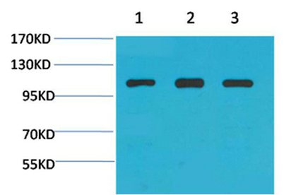
| Observed band (KD) | 103kD |
| Background | Alpha actinins belong to the spectrin gene superfamily which represents a diverse group of cytoskeletal proteins, including the alpha and beta spectrins and dystrophins. Alpha actinin is an actin-binding protein with multiple roles in different cell types. In nonmuscle cells, the cytoskeletal isoform is found along microfilament bundles and adherens-type junctions, where it is involved in binding actin to the membrane. In contrast, skeletal, cardiac, and smooth muscle isoforms are localized to the Z-disc and analogous dense bodies, where they help anchor the myofibrillar actin filaments. This gene encodes a nonmuscle, cytoskeletal, alpha actinin isoform and maps to the same site as the structurally similar erythroid beta spectrin gene. Three transcript variants encoding different isoforms have been found for this gene. [provided by RefSeq, Jul 2008], |
| Function | function:F-actin cross-linking protein which is thought to anchor actin to a variety of intracellular structures. This is a bundling protein.,similarity:Belongs to the alpha-actinin family.,similarity:Contains 1 actin-binding domain.,similarity:Contains 2 CH (calponin-homology) domains.,similarity:Contains 2 EF-hand domains.,similarity:Contains 4 spectrin repeats.,subcellular location:Colocalizes with MYOZ2 and PPP3CA at the Z-line of heart and skeletal muscle.,subunit:Homodimer; antiparallel. Interacts with DDN, MYOZ2, PDLIM2, TTID and LPP., |
| Subcellular location | Cytoplasm, cytoskeleton . Cytoplasm, myofibril, sarcomere, Z line . Cell membrane . Cell junction . Cell projection, ruffle . Colocalizes with MYOZ2 and PPP3CA at the Z-line of heart and skeletal muscle. Colocalizes with PSD in membrane ruffles and central reticular structures. . |
| Expression | Colon,Epithelium,Hepatocyte,Neuroblastoma,Placenta,Platelet,Skin,Tumor of tongue, |

Use collapsible tabs for more detailed information that will help customers make a purchasing decision.
Ex: Shipping and return policies, size guides, and other common questions.