 Français
 English
Cyclin E1 (phospho Thr77) rabbit pAb
AO-06-ES7919-50

| ELK.No | ES7919 |
| Product name | Cyclin E1 (phospho Thr77) rabbit pAb |
| Reactivity | Human;Rat;Mouse; |
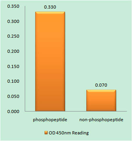
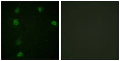
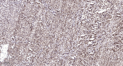
| Applications | IHC;IF;ELISA |
| Other name | CCNE1; CCNE; G1/S-specific cyclin-E1 |
| Size | 50μL |
| Unit price ($) | 148 |
| Human gene ID | 898 |
| Human Swiss-Prot | P24864 |
| Source | Rabbit |
| Isotype | IgG |
| Target | Cyclin E1 |
| Fields | >>Cell cycle;>>Oocyte meiosis;>>p53 signaling pathway;>>PI3K-Akt signaling pathway;>>Cellular senescence;>>Cushing syndrome;>>Hepatitis B;>>Measles;>>Human papillomavirus infection;>>Human T-cell leukemia virus 1 infection;>>Epstein-Barr virus infection;>>Pathways in cancer;>>Viral carcinogenesis;>>MicroRNAs in cancer;>>Prostate cancer;>>Small cell lung cancer;>>Gastric cancer |
| Gene name | CCNE1 |
| Protein name | G1/S-specific cyclin-E1 |
| Human gene link | View Human Gene |
| Human Swiss link | View Human Swiss-Prot |
| Mouse gene ID | |
| Mouse gene link | |
| Mouse Swiss-Prot | Q61457 |
| Mouse Swiss link | View Mouse Swiss-Prot |
| Rat gene ID | |
| Rat gene link | |
| Rat Swiss-Prot | |
| Rat Swiss link | |
| Immunogen | The antiserum was produced against synthesized peptide derived from human Cyclin E1 around the phosphorylation site of Thr77. AA range:43-92 |
| Specificity | Phospho-Cyclin E1 (T77) Polyclonal Antibody detects endogenous levels of Cyclin E1 protein only when phosphorylated at T77. |
| Formulation | Liquid in PBS containing 50% glycerol, 0.5% BSA and 0.02% sodium azide. |
| Clonality | Polyclonal |
| Dilution | Immunohistochemistry: 1/100 - 1/300. Immunofluorescence: 1/200 - 1/1000. ELISA: 1/10000. Not yet tested in other applications. |
| Purification | The antibody was affinity-purified from rabbit antiserum by affinity-chromatography using epitope-specific immunogen. |
| Concentration | 1 mg/ml |
| Storage stability | -20°C/1 year |
| Molecular Weight (Da) | 47kD |
| Observed band (KD) | |
| Background | The protein encoded by this gene belongs to the highly conserved cyclin family, whose members are characterized by a dramatic periodicity in protein abundance through the cell cycle. Cyclins function as regulators of CDK kinases. Different cyclins exhibit distinct expression and degradation patterns which contribute to the temporal coordination of each mitotic event. This cyclin forms a complex with and functions as a regulatory subunit of CDK2, whose activity is required for cell cycle G1/S transition. This protein accumulates at the G1-S phase boundary and is degraded as cells progress through S phase. Overexpression of this gene has been observed in many tumors, which results in chromosome instability, and thus may contribute to tumorigenesis. This protein was found to associate with, and be involved in, the phosphorylation of NPAT protein (nuclear protein mapped to the ATM locus), which participates in |
| Function | function:Essential for the control of the cell cycle at the G1/S (start) transition.,PTM:Phosphorylation of Thr-395 by GSK3 and of Ser-399 by CDK2 accelerates degradation via the ubiquitin proteasome pathway. Phosphorylated upon DNA damage, probably by ATM or ATR.,similarity:Belongs to the cyclin family. Cyclin E subfamily.,subunit:Interacts with a member of the CDK2/CDK protein kinases to form a serine/threonine kinase holoenzyme complex. The cyclin subunit imparts substrate specificity to the complex. Interacts with retinoblastoma binding protein 3 and retinoblastoma-like protein 1. Found in a complex with CDK2, CABLES1 and CCNA1 (By similarity). Part of a complex consisting of UHRF2, CDK2 and CCNE1.,tissue specificity:Highly expressed in testis and placenta. Low levels in bronchial epithelial cells., |
| Subcellular location | Nucleus . |
| Expression | Highly expressed in testis and placenta. Low levels in bronchial epithelial cells. |

Use collapsible tabs for more detailed information that will help customers make a purchasing decision.
Ex: Shipping and return policies, size guides, and other common questions.