 Français
 English
CD4 (phospho Ser433) rabbit pAb
AO-06-ES7987-100

| ELK.No | ES7987 |
| Product name | CD4 (phospho Ser433) rabbit pAb |
| Reactivity | Human;Mouse |
| Applications | IHC;IF;ELISA |
| Other name | CD4; T-cell surface glycoprotein CD4; T-cell surface antigen T4/Leu-3; CD antigen CD4 |
| Size | 100μL |
| Unit price ($) | 248 |
| Human gene ID | 920 |
| Human Swiss-Prot | P01730 |
| Source | Rabbit |
| Isotype | IgG |
| Target | CD4 |
| Fields | >>Viral life cycle - HIV-1;>>Cytokine-cytokine receptor interaction;>>Cell adhesion molecules;>>Antigen processing and presentation;>>Hematopoietic cell lineage;>>Th1 and Th2 cell differentiation;>>Th17 cell differentiation;>>T cell receptor signaling pathway;>>Yersinia infection;>>Human T-cell leukemia virus 1 infection;>>Human immunodeficiency virus 1 infection;>>PD-L1 expression and PD-1 checkpoint pathway in cancer;>>Primary immunodeficiency |
| Gene name | CD4 |
| Protein name | T-cell surface glycoprotein CD4 |
| Human gene link | View Human Gene |
| Human Swiss link | View Human Swiss-Prot |
| Mouse gene ID | 12504 |
| Mouse gene link | View Mouse Gene |
| Mouse Swiss-Prot | P06332 |
| Mouse Swiss link | View Mouse Swiss-Prot |
| Rat gene ID | |
| Rat gene link | |
| Rat Swiss-Prot | |
| Rat Swiss link | |
| Immunogen | The antiserum was produced against synthesized peptide derived from human CD4 around the phosphorylation site of Ser433. AA range:401-450 |
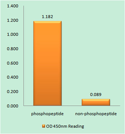
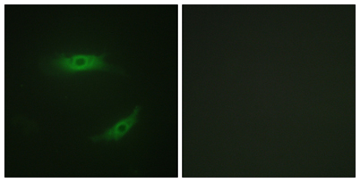
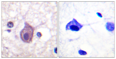
| Specificity | Phospho-CD4 (S433) Polyclonal Antibody detects endogenous levels of CD4 protein only when phosphorylated at S433. |
| Formulation | Liquid in PBS containing 50% glycerol, 0.5% BSA and 0.02% sodium azide. |
| Clonality | Polyclonal |
| Dilution | Immunohistochemistry: 1/100 - 1/300. Immunofluorescence: 1/200 - 1/1000. ELISA: 1/40000. Not yet tested in other applications. |
| Purification | The antibody was affinity-purified from rabbit antiserum by affinity-chromatography using epitope-specific immunogen. |
| Concentration | 1 mg/ml |
| Storage stability | -20°C/1 year |
| Molecular Weight (Da) | 51kD |
| Observed band (KD) | |
| Background | This gene encodes a membrane glycoprotein of T lymphocytes that interacts with major histocompatibility complex class II antigenes and is also a receptor for the human immunodeficiency virus. This gene is expressed not only in T lymphocytes, but also in B cells, macrophages, and granulocytes. It is also expressed in specific regions of the brain. The protein functions to initiate or augment the early phase of T-cell activation, and may function as an important mediator of indirect neuronal damage in infectious and immune-mediated diseases of the central nervous system. Multiple alternatively spliced transcript variants encoding different isoforms have been identified in this gene. [provided by RefSeq, Aug 2010], |
| Function | function:Accessory protein for MHC class-II antigen/T-cell receptor interaction. May regulate T-cell activation. Induces the aggregation of lipid rafts.,miscellaneous:Primary receptor for HIV-1.,online information:CD4 entry,PTM:Palmitoylation and association with LCK contribute to the enrichment of CD4 in lipid rafts.,similarity:Contains 1 Ig-like V-type (immunoglobulin-like) domain.,similarity:Contains 3 Ig-like C2-type (immunoglobulin-like) domains.,subcellular location:Localizes to lipid rafts. Removed from plasma membrane by HIV-1 Nef protein that increases clathrin-dependent endocytosis of this antigen to target it to lysosomal degradation. Cell surface expression is also down-modulated by HIV-1 Envelope polyprotein gp160 that interacts with, and sequesters CD4 in the endoplasmic reticulum.,subunit:Associates with LCK. Binds to HIV-1 gp120 and to P4HB/PDI and upon HIV-1 binding to t |
| Subcellular location | Cell membrane ; Single-pass type I membrane protein . Localizes to lipid rafts (PubMed:12517957, PubMed:9168119). Removed from plasma membrane by HIV-1 Nef protein that increases clathrin-dependent endocytosis of this antigen to target it to lysosomal degradation. Cell surface expression is also down-modulated by HIV-1 Envelope polyprotein gp160 that interacts with, and sequesters CD4 in the endoplasmic reticulum. |
| Expression | Highly expressed in T-helper cells. The presence of CD4 is a hallmark of T-helper cells which are specialized in the activation and growth of cytotoxic T-cells, regulation of B cells, or activation of phagocytes. CD4 is also present in other immune cells such as macrophages, dendritic cells or NK cells. |

Use collapsible tabs for more detailed information that will help customers make a purchasing decision.
Ex: Shipping and return policies, size guides, and other common questions.