 Français
 English
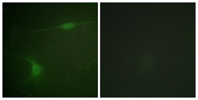
JIP-1 (phospho Thr103) rabbit pAb
AO-06-ES8048-50

| ELK.No | ES8048 |
| Product name | JIP-1 (phospho Thr103) rabbit pAb |
| Reactivity | Human;Mouse;Rat |
| Applications | WB;IHC;IF;ELISA |
| Other name | MAPK8IP1; IB1; JIP1; PRKM8IP; C-Jun-amino-terminal kinase-interacting protein 1; JIP-1; JNK-interacting protein 1; Islet-brain 1; IB-1; JNK MAP kinase scaffold protein 1; Mitogen-activated protein kinase 8-interacting protein 1 |
| Size | 50μL |
| Unit price ($) | 148 |
| Human gene ID | 9479 |
| Human Swiss-Prot | Q9UQF2 |
| Source | Rabbit |
| Isotype | IgG |
| Target | JIP-1 |
| Fields | >>MAPK signaling pathway |
| Gene name | MAPK8IP1 |
| Protein name | C-Jun-amino-terminal kinase-interacting protein 1 |
| Human gene link | View Human Gene |
| Human Swiss link | View Human Swiss-Prot |
| Mouse gene ID | 19099 |
| Mouse gene link | View Mouse Gene |
| Mouse Swiss-Prot | Q9WVI9 |
| Mouse Swiss link | View Mouse Swiss-Prot |
| Rat gene ID | 116457 |
| Rat gene link | View Rat Gene |
| Rat Swiss-Prot | Q9R237 |
| Rat Swiss link | View Rat Swiss-Prot |
| Immunogen | The antiserum was produced against synthesized peptide derived from human JIP1 around the phosphorylation site of Thr103. AA range:69-118 |
| Specificity | Phospho-JIP-1 (T103) Polyclonal Antibody detects endogenous levels of JIP-1 protein only when phosphorylated at T103. |
| Formulation | Liquid in PBS containing 50% glycerol, 0.5% BSA and 0.02% sodium azide. |
| Clonality | Polyclonal |
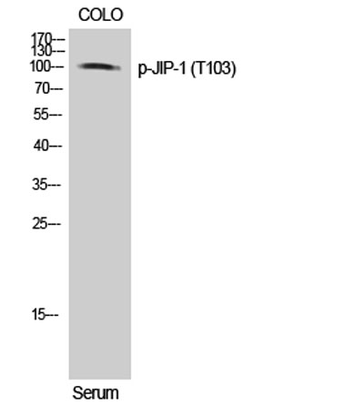
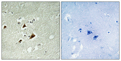
| Dilution | Western Blot: 1/500 - 1/2000. Immunohistochemistry: 1/100 - 1/300. Immunofluorescence: 1/200 - 1/1000. ELISA: 1/5000. Not yet tested in other applications. |
| Purification | The antibody was affinity-purified from rabbit antiserum by affinity-chromatography using epitope-specific immunogen. |
| Concentration | 1 mg/ml |
| Storage stability | -20°C/1 year |
| Molecular Weight (Da) | |
| Observed band (KD) | 113kD |
| Background | This gene encodes a regulator of the pancreatic beta-cell function. It is highly similar to JIP-1, a mouse protein known to be a regulator of c-Jun amino-terminal kinase (Mapk8). This protein has been shown to prevent MAPK8 mediated activation of transcription factors, and to decrease IL-1 beta and MAP kinase kinase 1 (MEKK1) induced apoptosis in pancreatic beta cells. This protein also functions as a DNA-binding transactivator of the glucose transporter GLUT2. RE1-silencing transcription factor (REST) is reported to repress the expression of this gene in insulin-secreting beta cells. This gene is found to be mutated in a type 2 diabetes family, and thus is thought to be a susceptibility gene for type 2 diabetes. [provided by RefSeq, May 2011], |
| Function | disease:Defects in MAPK8IP1 are a cause of non-insulin-dependent diabetes mellitus (NIDDM) [MIM:125853]. NIDDM is characterized by an autosomal dominant mode of inheritance, onset during adulthood and insulin resistance.,domain:A minimal inhibitory domain prevents pancreatic beta cell apoptosis in vitro, and prevents activation of c-jun by MAPK8, MAPK9 and MAPK10.,domain:The destruction boxes (D-box) may act as recognition signals for degradation via the ubiquitin-proteasome pathway.,function:The JNK-interacting protein (JIP) group of scaffold proteins selectively mediates JNK signaling by aggregating specific components of the MAPK cascade to form a functional JNK signaling module. Required for JNK activation in response to excitotoxic stress. Cytoplasmic MAPK8IP1 causes inhibition of JNK-regulated activity by retaining JNK in the cytoplasm and inhibiting JNK phosphorylation of c-Jun. M |
| Subcellular location | Cytoplasm . Cytoplasm, perinuclear region . Nucleus . Endoplasmic reticulum membrane. Mitochondrion membrane. Accumulates in cell surface projections. Under certain stress conditions, translocates to the perinuclear region of neurons. In insulin-secreting cells, detected in both the cytoplasm and nucleus (By similarity). . |
| Expression | Highly expressed in brain. Expressed in neurons, localizing to neurite tips in differentiating cells. Also expressed in the pancreas, testis and prostate. Low levels in heart, ovary and small intestine. Decreased levels in pancreatic beta cells sensitize cells to IL-1-beta-induced apoptosis. |

Use collapsible tabs for more detailed information that will help customers make a purchasing decision.
Ex: Shipping and return policies, size guides, and other common questions.