 Français
 English
CD40 (phospho Thr254) rabbit pAb
AO-06-ES8067-100

| ELK.No | ES8067 |
| Product name | CD40 (phospho Thr254) rabbit pAb |
| Reactivity | Human;Mouse |
| Applications | WB;ELISA |
| Other name | CD40; TNFRSF5; Tumor necrosis factor receptor superfamily member 5; B-cell surface antigen CD40; Bp50; CD40L receptor; CDw40; CD antigen CD40 |
| Size | 100μL |
| Unit price ($) | 248 |
| Human gene ID | 958 |
| Human Swiss-Prot | P25942 |
| Source | Rabbit |
| Isotype | IgG |
| Target | CD40 |
| Fields | >>Cytokine-cytokine receptor interaction;>>NF-kappa B signaling pathway;>>Cell adhesion molecules;>>Toll-like receptor signaling pathway;>>Intestinal immune network for IgA production;>>Malaria;>>Toxoplasmosis;>>Human T-cell leukemia virus 1 infection;>>Epstein-Barr virus infection;>>Transcriptional misregulation in cancer;>>Asthma;>>Autoimmune thyroid disease;>>Systemic lupus erythematosus;>>Allograft rejection;>>Primary immunodeficiency;>>Viral myocarditis;>>Lipid and atherosclerosis |
| Gene name | CD40 |
| Protein name | Tumor necrosis factor receptor superfamily member 5 |
| Human gene link | View Human Gene |
| Human Swiss link | View Human Swiss-Prot |
| Mouse gene ID | 21939 |
| Mouse gene link | View Mouse Gene |
| Mouse Swiss-Prot | P27512 |
| Mouse Swiss link | View Mouse Swiss-Prot |
| Rat gene ID | |
| Rat gene link | |
| Rat Swiss-Prot | |
| Rat Swiss link | |
| Immunogen | The antiserum was produced against synthesized peptide derived from human TNFRSF5 around the phosphorylation site of Thr254. AA range:220-269 |
| Specificity | Phospho-CD40 (T254) Polyclonal Antibody detects endogenous levels of CD40 protein only when phosphorylated at T254. |
| Formulation | Liquid in PBS containing 50% glycerol, 0.5% BSA and 0.02% sodium azide. |
| Clonality | Polyclonal |
| Dilution | Western Blot: 1/500 - 1/2000. ELISA: 1/5000. Not yet tested in other applications. |
| Purification | The antibody was affinity-purified from rabbit antiserum by affinity-chromatography using epitope-specific immunogen. |
| Concentration | 1 mg/ml |
| Storage stability | -20°C/1 year |
| Molecular Weight (Da) | |
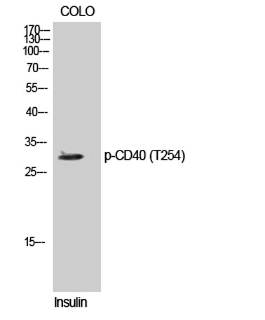
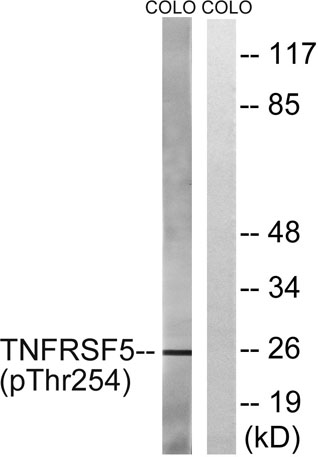
| Observed band (KD) | 30kD |
| Background | This gene is a member of the TNF-receptor superfamily. The encoded protein is a receptor on antigen-presenting cells of the immune system and is essential for mediating a broad variety of immune and inflammatory responses including T cell-dependent immunoglobulin class switching, memory B cell development, and germinal center formation. AT-hook transcription factor AKNA is reported to coordinately regulate the expression of this receptor and its ligand, which may be important for homotypic cell interactions. Adaptor protein TNFR2 interacts with this receptor and serves as a mediator of the signal transduction. The interaction of this receptor and its ligand is found to be necessary for amyloid-beta-induced microglial activation, and thus is thought to be an early event in Alzheimer disease pathogenesis. Mutations affecting this gene are the cause of autosomal recessive hyper-IgM immunodeficiency type 3 (HIG |
| Function | alternative products:Additional isoforms seem to exist,disease:Defects in CD40 are the cause of hyper-IgM immunodeficiency type 3 (HIGM3) [MIM:606843]. HIGM3 is an autosomal recessive disorder which includes an inability of B cells to undergo isotype switching, one of the final differentiation steps in the humoral immune system, an inability to mount an antibody-specific immune response, and a lack of germinal center formation.,function:Receptor for TNFSF5/CD40LG.,online information:CD40 entry,online information:CD40 mutation db,similarity:Contains 4 TNFR-Cys repeats.,subunit:Monomer and homodimer. The variant form found in the bladder carcinoma cell line Hu549 does not form homodimers. Interacts with TRAF1, TRAF2, TRAF3, TRAF5 and TRAF6.,tissue specificity:B-cells and in primary carcinomas., |
| Subcellular location | [Isoform I]: Cell membrane; Single-pass type I membrane protein.; [Isoform II]: Secreted. |
| Expression | B-cells and in primary carcinomas. |

Use collapsible tabs for more detailed information that will help customers make a purchasing decision.
Ex: Shipping and return policies, size guides, and other common questions.