 Français
 English

| ELK.No | ES8120 |
| Product name | TBC1D4 rabbit pAb |
| Reactivity | Human;Mouse |
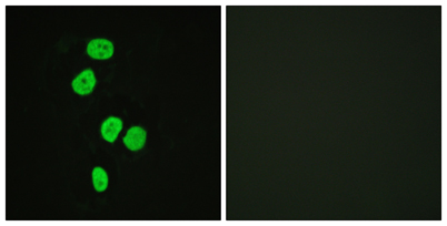
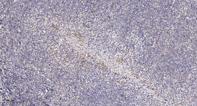
| Applications | IHC;IF;ELISA |
| Other name | TBC1D4; AS160; KIAA0603; TBC1 domain family member 4; Akt substrate of 160 kDa; AS160 |
| Size | 50μL |
| Unit price ($) | 148 |
| Human gene ID | 9882 |
| Human Swiss-Prot | O60343 |
| Source | Rabbit |
| Isotype | IgG |
| Target | TBC1D4 |
| Fields | >>Thyroid hormone signaling pathway;>>Insulin resistance;>>Diabetic cardiomyopathy |
| Gene name | TBC1D4 |
| Protein name | TBC1 domain family member 4 |
| Human gene link | View Human Gene |
| Human Swiss link | View Human Swiss-Prot |
| Mouse gene ID | 210789 |
| Mouse gene link | View Mouse Gene |
| Mouse Swiss-Prot | Q8BYJ6 |
| Mouse Swiss link | View Mouse Swiss-Prot |
| Rat gene ID | |
| Rat gene link | |
| Rat Swiss-Prot | |
| Rat Swiss link | |
| Immunogen | The antiserum was produced against synthesized peptide derived from human AS160. AA range:611-660 |
| Specificity | TBC1D4 Polyclonal Antibody detects endogenous levels of TBC1D4 protein. |
| Formulation | Liquid in PBS containing 50% glycerol, 0.5% BSA and 0.02% sodium azide. |
| Clonality | Polyclonal |
| Dilution | Immunohistochemistry: 1/100 - 1/300. Immunofluorescence: 1/200 - 1/1000. ELISA: 1/10000. Not yet tested in other applications. |
| Purification | The antibody was affinity-purified from rabbit antiserum by affinity-chromatography using epitope-specific immunogen. |
| Concentration | 1 mg/ml |
| Storage stability | -20°C/1 year |
| Molecular Weight (Da) | 147kD |
| Observed band (KD) | |
| Background | This gene is a member of the Tre-2/BUB2/CDC16 domain family. The protein encoded by this gene is a Rab-GTPase-activating protein, and contains two phopshotyrosine-binding domains (PTB1 and PTB2), a calmodulin-binding domain (CBD), a Rab-GTPase domain, and multiple AKT phosphomotifs. This protein is thought to play an important role in glucose homeostasis by regulating the insulin-dependent trafficking of the glucose transporter 4 (GLUT4), important for removing glucose from the bloodstream into skeletal muscle and fat tissues. Reduced expression of this gene results in an increase in GLUT4 levels at the plasma membrane, suggesting that this protein is important in intracellular retention of GLUT4 under basal conditions. When exposed to insulin, this protein is phosphorylated, dissociates from GLUT4 vesicles, resulting in increased GLUT4 at the cell surface, and enhanced glucose transport. Ph |
| Function | disease:May be involved in atopic dermatitis (AD).,function:May act as a GTPase-activating protein for RAB2A, RAB8A, RAB10 and RAB14. Isoform 2 promotes insulin-induced glucose transporter SLC2A4/GLUT4 translocation at the plasma membrane, thus increasing glucose uptake.,PTM:Insulin-stimulated phosphorylation is required for SLC2A4/GLUT4 translocation.,PTM:Phosphorylated by AKT1; insulin-induced.,PTM:Physiological hyperinsulinemia increases phosphorylation in skeletal muscle. Insulin-stimulated phosphorylation is reduced by 39% in type 2 diabetic patients.,similarity:Contains 1 Rab-GAP TBC domain.,similarity:Contains 2 PID domains.,subcellular location:Isoform 2 shows a cytoplasmic perinuclear localization in a myoblastic cell line in resting and insulin-stimulated cells.,tissue specificity:Widely expressed, but differential expression for isoforms 1 and 2, with highest overall expressio |
| Subcellular location | Cytoplasm . Isoform 2 shows a cytoplasmic perinuclear localization in a myoblastic cell line in resting and insulin-stimulated cells. |
| Expression | Widely expressed. Isoform 2 is the highest overexpressed in most tissues. Isoform 1 is highly expressed in skeletal muscle and heart, but was not detectable in the liver nor in adipose tissue. Isoform 2 is strongly expressed in adrenal and thyroid gland, and also in lung, kidney, colon, brain and adipose tissue. Isoform 2 is moderately expressed in skeletal muscle. Expressed in pancreatic Langerhans islets, including beta cells (at protein level). Expression is decreased by twofold in pancreatic islets in type 2 diabetes patients compared to control subjects. Up-regulated in T-cells from patients with atopic dermatitis. |

Use collapsible tabs for more detailed information that will help customers make a purchasing decision.
Ex: Shipping and return policies, size guides, and other common questions.