 Français
 English

| ELK.No | ES8387 |
| Product name | CD26 rabbit pAb |
| Reactivity | Human;Rat;Mouse; |
| Applications | WB;IHC;IF;ELISA |
| Other name | DPP4; ADCP2; CD26; Dipeptidyl peptidase 4; ADABP; Adenosine deaminase complexing protein 2; ADCP-2; Dipeptidyl peptidase IV; DPP IV; T-cell activation antigen CD26; TP103; CD26 |
| Size | 100μL |
| Unit price ($) | 248 |
| Human gene ID | 1803 |
| Human Swiss-Prot | P27487 |
| Source | Rabbit |
| Isotype | IgG |
| Target | CD26 |
| Fields | >>Protein digestion and absorption |
| Gene name | DPP4 |
| Protein name | Dipeptidyl peptidase 4 |
| Human gene link | View Human Gene |
| Human Swiss link | View Human Swiss-Prot |
| Mouse gene ID | |
| Mouse gene link | |
| Mouse Swiss-Prot | P28843 |
| Mouse Swiss link | View Mouse Swiss-Prot |
| Rat gene ID | |
| Rat gene link | |
| Rat Swiss-Prot | |
| Rat Swiss link | |
| Immunogen | The antiserum was produced against synthesized peptide derived from the Internal region of human DPP4. AA range:350-400 |
| Specificity | CD26 Polyclonal Antibody detects endogenous levels of CD26 protein. |
| Formulation | Liquid in PBS containing 50% glycerol, 0.5% BSA and 0.02% sodium azide. |
| Clonality | Polyclonal |
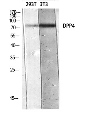
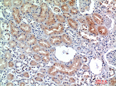
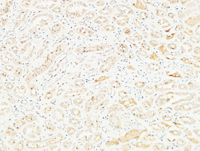
| Dilution | Western Blot: 1/500 - 1/2000. IHC-p: 1:100-1:300. ELISA: 1/10000. Not yet tested in other applications. |
| Purification | The antibody was affinity-purified from rabbit antiserum by affinity-chromatography using epitope-specific immunogen. |
| Concentration | 1 mg/ml |
| Storage stability | -20°C/1 year |
| Molecular Weight (Da) | |
| Observed band (KD) | 105kD |
| Background | The protein encoded by this gene is identical to adenosine deaminase complexing protein-2, and to the T-cell activation antigen CD26. It is an intrinsic membrane glycoprotein and a serine exopeptidase that cleaves X-proline dipeptides from the N-terminus of polypeptides. [provided by RefSeq, Jul 2008], |
| Function | catalytic activity:Release of an N-terminal dipeptide, Xaa-Yaa-|-Zaa-, from a polypeptide, preferentially when Yaa is Pro, provided Zaa is neither Pro nor hydroxyproline.,function:Removes N-terminal dipeptides sequentially from polypeptides having unsubstituted N-termini provided that the penultimate residue is proline. Plays a role in T-cell activation.,online information:Dipeptidyl peptidase-4 entry,PTM:The soluble form (SDPP) derives from the membrane form (MDPP) by proteolytic processing.,similarity:Belongs to the peptidase S9B family. DPPIV subfamily.,subunit:Homodimer or heterodimer with Seprase (FAP).,tissue specificity:Expressed in the poorly differentiated crypt cells of the small intestine as well as in the mature villous cells. Expressed at very low levels in the colon., |
| Subcellular location | [Dipeptidyl peptidase 4 soluble form]: Secreted . Detected in the serum and the seminal fluid. .; Cell membrane ; Single-pass type II membrane protein. Apical cell membrane ; Single-pass type II membrane protein. Cell projection, invadopodium membrane ; Single-pass type II membrane protein. Cell projection, lamellipodium membrane ; Single-pass type II membrane protein. Cell junction . Membrane raft . Translocated to the apical membrane through the concerted action of N- and O-Glycans and its association with lipid microdomains containing cholesterol and sphingolipids (PubMed:11773049). Redistributed to membrane rafts in T-cell in an interleukin-12-dependent activation (PubMed:12676959). Its interaction with CAV1 is necessary for its translocation to membrane rafts (PubMed:17287217). Coloca |
| Expression | Expressed specifically in lymphatic vessels but not in blood vessels in the skin, small intestine, esophagus, ovary, breast and prostate glands. Not detected in lymphatic vessels in the lung, kidney, uterus, liver and stomach (at protein level). Expressed in the poorly differentiated crypt cells of the small intestine as well as in the mature villous cells. Expressed at very low levels in the colon. |

Use collapsible tabs for more detailed information that will help customers make a purchasing decision.
Ex: Shipping and return policies, size guides, and other common questions.